Resources
Case Studies
-
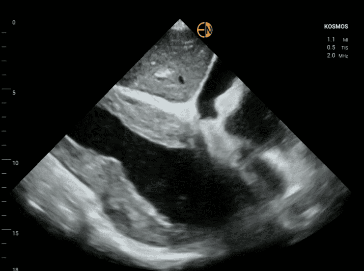
Abdominal Aortic Aneurysm
Abdominal Aortic Aneurysm Patient: A 63 year old male patient presented to the ER with peripheral artery embolization (blue toe syndrome of lower limbs). Initial Examination: He had a history of well controlled hypertension and long…
-

Aortic Dissection
Aortic Dissection A 55-year-old, obese (BMI 33 kg/m2) female patient with long-standing hypertension presented to the ER with chest pain radiating to the back. Her ECG had no signs of ischemia. Bedside examination with KOSMOS…
-
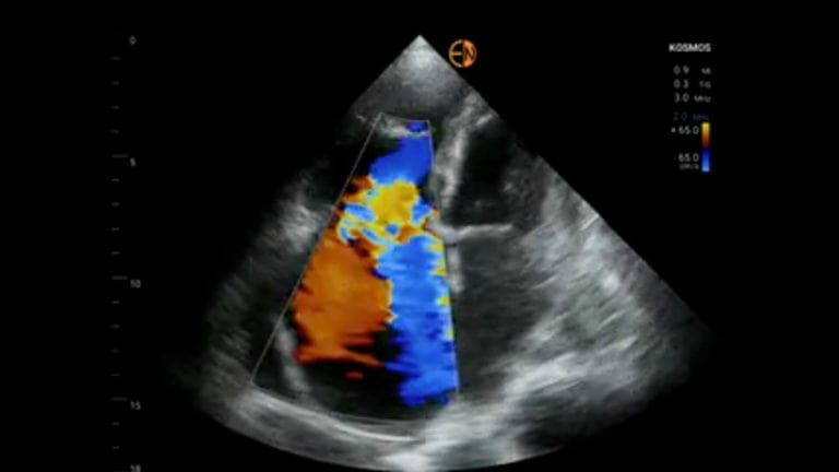
Finding Criteria for Myocardial Rupture With POCUS AI
Finding Criteria for Myocardial Rupture With POCUS AI A 75 year old female patient presented to the ER with anasarca edema. She had a history of permanent atrial fibrillation and hypertension. On clinical examination increased…
-
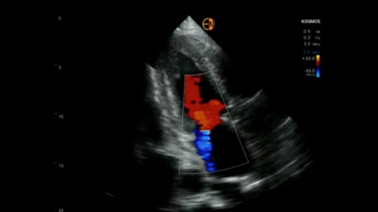
Amyloidosis Diagnosis With POCUS AI
Finding Criteria for Amyloidosis Diagnosis With POCUS AI A 73 year old male patient presented with right sided heart failure. His ECG revealed atrial fibrillation. On clinical examination increased jugular venous pressure and a holosystolic…
-
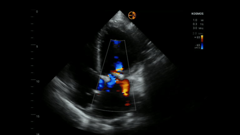
Identifying & Diagnosing Ischemic Mitral Regurgitation
Identifying & Diagnosing Ischemic Mitral Regurgitation Patient: A 51 year old female patient presented to the ER with gradually worsening shortness of breath. Initial Examination: She had a history of coronary artery disease and ischemic…
-
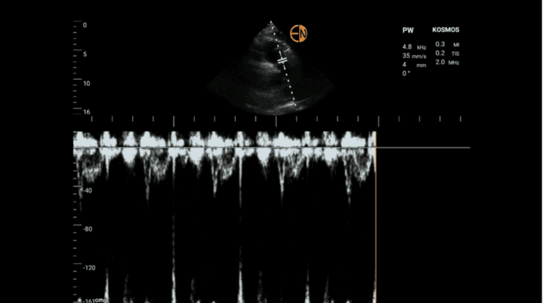
Identifying & Diagnosing a Pulmonary Embolism
Identifying & Diagnosing a Pulmonary Embolism Initial Examination: He reported SOBOE during the last three days. On clinical examination increased jugular venous pressure and low systolic blood pressure was noticed. Kosmos POCUS Examination: Bedside examination…
-

Identifying & Diagnosing Aortic Stenosis
Identifying & Diagnosing Aortic Stenosis Patient: An 82 year old female patient presented to the ER with symptoms and signs of congestive heart failure. Initial Examination: She had a history of well controlled hypertension treated…
-
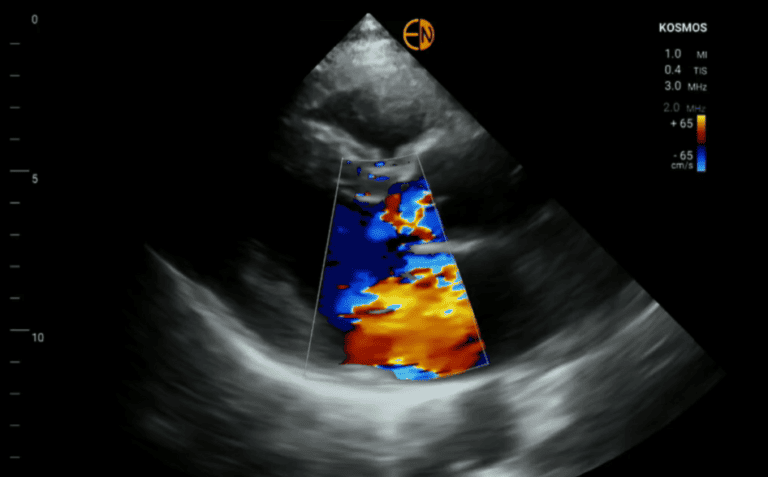
Mitral Valve Fibroelastoma via Echocardiogram
Diagnosing Mitral Valve Fibroelastoma via Echocardiogram Patient: A 53-year-old male patient with no previous medical history presented to the ER with TIA symptoms. Initial Examination: An echocardiogram was requested to exclude cardiac causes of embolization.…
-
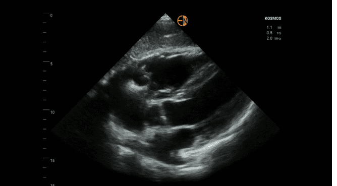
Finding Criteria for Arrhythmogenic Right Ventricular Cardiomyopathy (ARVC) with Ultrasound
Finding Criteria for Arrhythmogenic Right Ventricular Cardiomyopathy (ARVC) with Ultrasound AI Station, CompanyDecember 2, 2021 Patient: A 59-year-old male patient with no previous medical history presented to the ER with dizziness. Initial Examination: The patient…