Recent Posts
-
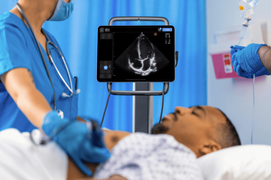
Cardiac Ultrasound Machines in 2026: Why the Best Choice Isn’t Necessarily a Traditional Cart
Cardiac Ultrasound Machines in 2026: Why the Best Choice Isn’t Necessarily a Traditional Cart For most of the last two decades, a cardiac ultrasound machine meant more or less one thing: a cart in the…
-
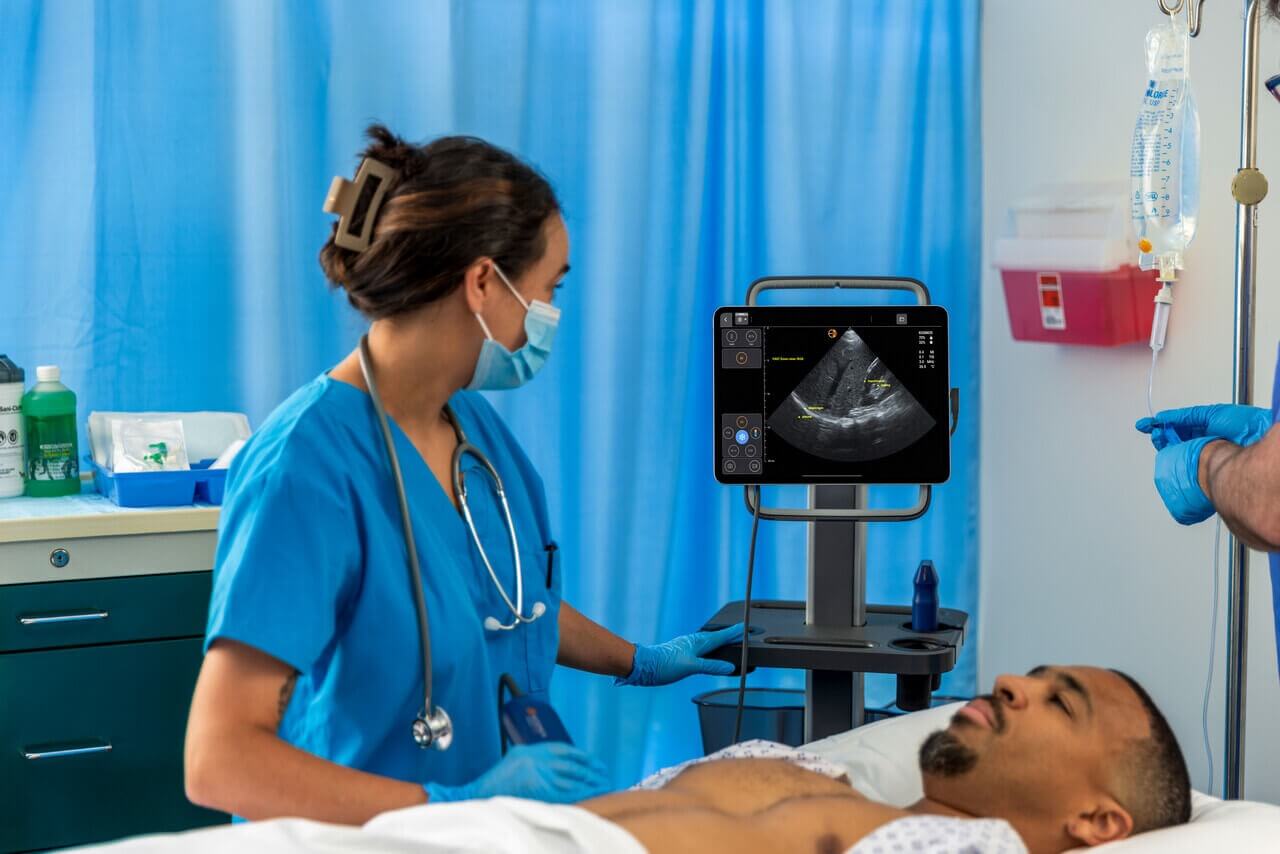
Advancing Perioperative Ultrasound: Dr. Deepak Borde on AI and Echoliteracy
Advancing Perioperative Ultrasound: Dr. Deepak Borde on AI and Echoliteracy Cardiac anesthesiologists rely on rapid access to accurate patient data. As point-of-care ultrasound (POCUS) evolves, the ability to perform complex assessments at the bedside is…
-

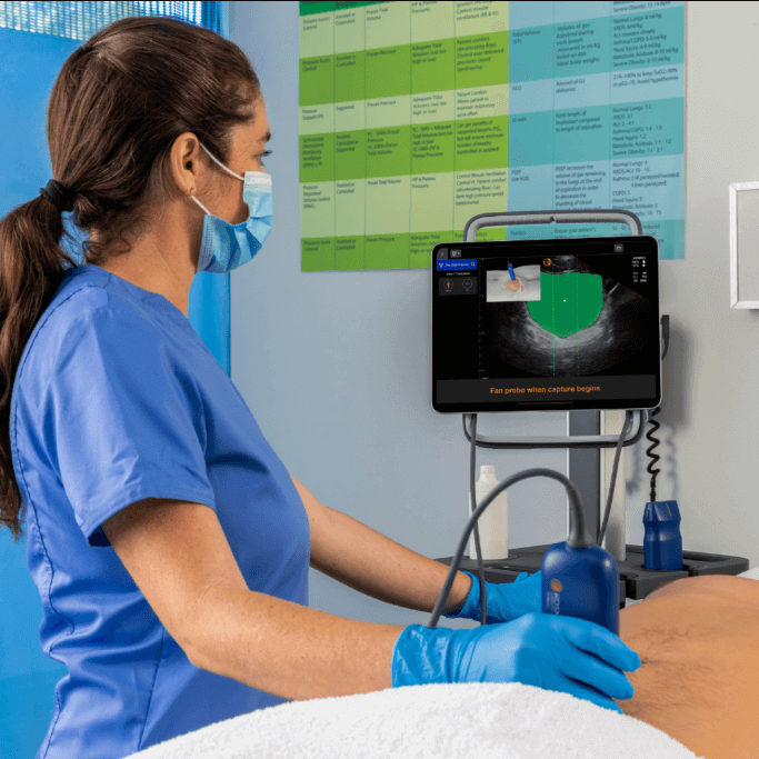
EchoNous Expands Kosmos Clinical Capabilities with Vascular Auto Flow; Advanced Cardiac and Connectivity Tools
EchoNous Expands Kosmos Clinical Capabilities with Vascular Auto Flow; Advanced Cardiac and Connectivity Tools REDMOND, Wash. — June 2026 — EchoNous, a leader in point-of-care ultrasound (POCUS) innovation, today announced a new release for the…
-
The Best Bladder Scanners in 2026: Comparison, Reviews, & Buyer’s Guide
The Best Bladder Scanners in 2026: Comparison, Reviews, & Buyer’s Guide Choosing a bladder scanner isn’t as complicated as it first appears, and it’s also not as simple as picking the most familiar brand or…