Diagnosing Mitral Valve Fibroelastoma via Echocardiogram
Patient: A 53-year-old male patient with no previous medical history presented to the ER with TIA symptoms.
Initial Examination: An echocardiogram was requested to exclude cardiac causes of embolization.
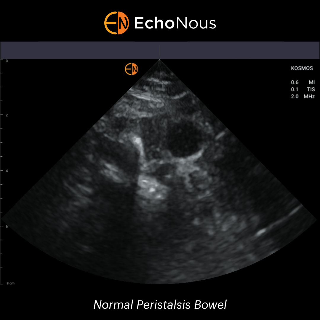
Kosmos POCUS Examination: Bedside cardiac examination with KOSMOS demonstrated bi-leaflet prolapse of the mitral valve with moderate regurgitation. Closer inspection of the valve revealed a small mobile mass on the anterior mitral valve leaflet which later proved to be a fibroelastoma (infective and non-bacterial thrombotic endocarditis were excluded with lab testing and the patient was taken for surgery).


