Identifying & Diagnosing a Pulmonary Embolism
Initial Examination: He reported SOBOE during the last three days. On clinical examination increased jugular venous pressure and low systolic blood pressure was noticed.
Kosmos POCUS Examination: Bedside examination with Kosmos revealed dilated right ventricle with positive McConnell sign, reduced systolic function (RV – PA uncoupling) and reduced RV stroke volume. The RA was also dilated. The LV was small in size and under filled. Systolic D-shape of the interventricular septum was noted indicating RV pressure overload. The IVC was dilated. All these features strongly pointed towards massive pulmonary embolism which was confirmed with urgent CTPA.
PLAX view showing dilated RV with impaired systolic function.
SAX view at the level of papillary muscles showing D-shape LV during systole due to RV pressure overload.
PW Doppler at the RVOT demonstrating reduced RV stroke volume and steep AcT due to increased afterload.
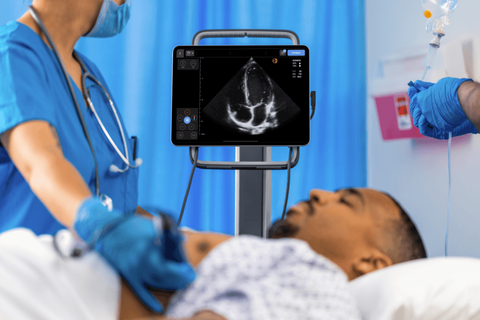
A4C view showing dilated RV with impaired systolic function (RV – PA uncoupling) and positive McConnell sign. The RA is also dilated.
Dilated IVC.