Teaching Cardiothoracic Anatomy with AI-Powered Ultrasound Systems
Aim
The study was designed to evaluate the effectiveness of an Artificial intelligence (A.I.) cardiothoracic ultrasound system (US system) as an anatomy educational tool and explore the possibility of a gender bias in the use of A.I. technology.
Introduction
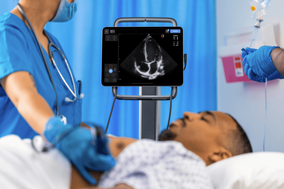
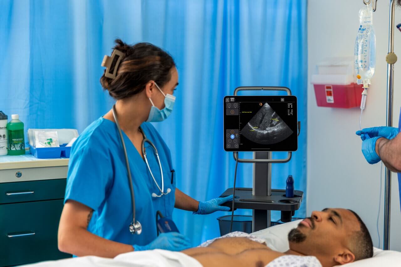
Introduction of A.I. enabled US systems to anatomy sessions has the potential to significantly affect medical education in the near future and enhance the capabilities of the next generation physicians.¹
In the last few years, A.I. has been introduced in medicine² with many applications in various fields like Radiology³, Cardiology⁴ and Neurology.⁵ It recently found its way into cardiothoracic ultrasound imaging systems used for Point of Care Ultrasound Examinations (POCUS) and has the potential to transform the clinical assessment of heart and lungs over the next decade.⁶
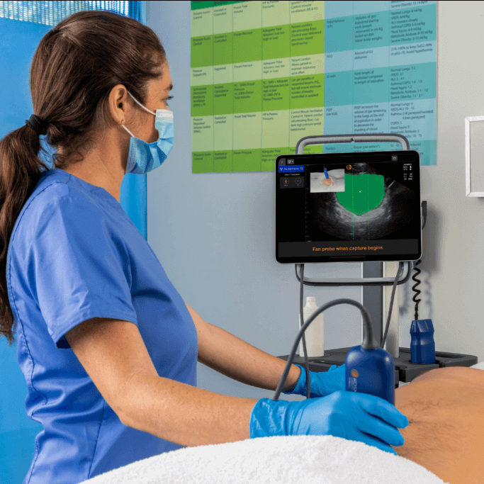
Ultrasound-based teaching is the cornerstone of living anatomy and offers the opportunity to medical students:
- to observe in real-time the movement of anatomical structures,
- to understand physiology and hemodynamics,
- to differentiate between normal and pathological variants, and
- to appreciate the relationship between surface and deep anatomy.
Using A.I. enabled POCUS to train the next generation of physicians will enable us to deliver better care to patients.⁷
It was hypothesized that A.I. enabled ultrasound teaching in an anatomy session will be an invaluable educational tool and that there is gender bias in the use of A.I. enabled technologies.
Methods
An analytical cross-sectional study was performed among 2nd year medical students (n=103) at the University of Nicosia Medical School, in Cyprus. Ethical approval of this study was obtained from the Cyprus National Bioethics Committee (CNBC).
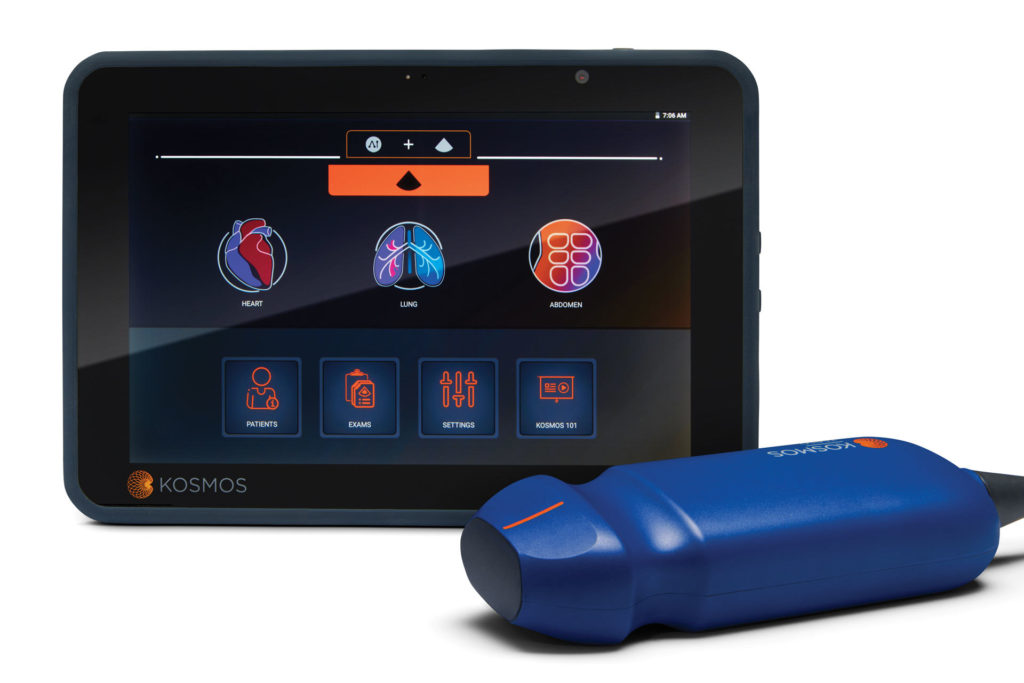
A questionnaire was distributed after the completion of a 20 minute anatomy lab practical that included a cardiology ultrasound session using the A.I. cardiothoracic ultrasound system.
Questionnaire asked students to rate: their experience on A.I. enabled ultrasound-based teaching and, the use of A.I. enabled ultrasound demonstrations in enhancing anatomy knowledge.
Analysis was done using SPSS version 26. The X2 test for comparison of binary and nominal variables was performed and statistical significance was determined as a p-value ≤0.05.
Results
Total of 103 responses were received from a cohort of 119 students since physical attendance was needed to complete the questionnaire. Participants consisted of 60 females and 43 males.
We aggregated male and female responses as there was no significant gender difference between the two in regards to the evaluation of the effectiveness of the A.I. system as an anatomy educational tool. However, both genders showed preference for the A.I. enabled ultrasound-based teaching method.
A question used to evaluate the project aim asked the participants to state if they believe that ultrasound demonstration with A.I. is a necessary adjunct to traditional teaching methods during anatomy labs. 97 responded positively with only 6 responding negatively.
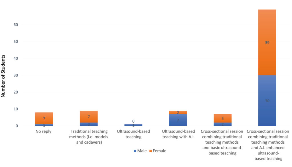
Key Finding: p-value = 0.05 was denoted with regards to which anatomy teaching method will be more beneficial to the participants’ anatomy knowledge. Figure 2 shows a significant gender bias in the choice of teaching method.
- 86% (n=37) of males preferred any teaching method involving A.I. in US systems when compared to 68% (n=41) of females.
Discussion
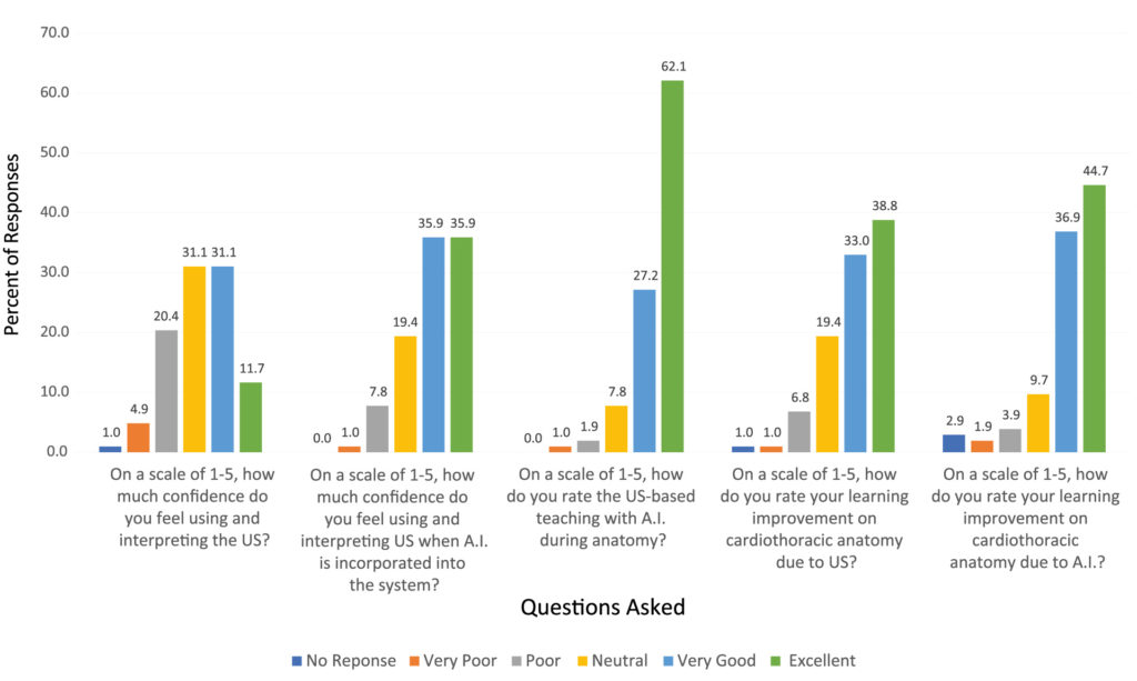
43% of the participants had excellent/very good confidence in using and interpreting US with no A.I. while with the A.I. incorporated the percentage increased by 29% to a value of 72%.
Moreover, 72% of participants rated their learning improvement due to ultrasounds as excellent/very good, while with A.I. incorporated there was a 12% difference with an increase to ≈ 84%.
The results indicate the confidence level and the improvement in anatomical knowledge of the students, had increased when the system had A.I. since it can provide immediate feedback and is in agreement with published literature of A.I. in medical education.⁹
94% of the responders want A.I. US demonstrations to be included as an addition to their traditional anatomy lab sessions. This finding is in line with recommendations that the medical curriculum should be reviewed to include A.I.¹
Key finding: A statistically significant gender bias is seen in the choice of teaching method with an 18% gender difference. As was reported by Denend et al.9 gender perception differences do exist in health technology where there is an increase in inhibition or initial operator exposure in females.
A limitation of this study is the introduction of selection bias as a result of the small sample size. Due to the study nature of self-reporting data, information bias can also be present.
Moreover, there was only one session delivered to the students with A.I. enabled US before they were asked to fill out the questionnaire.
Conclusions
- The majority of the students agreed that using A.I. enabled ultrasound systems in the anatomy labs was an improvement to the traditional teaching method.
- A.I. enabled ultrasounds increased students confidence in interpreting ultrasound images.
- Ultrasound-based teaching with A.I. during anatomy was highly appraised, and the students indicated that it had improved their cardiothoracic anatomy knowledge.
- This study, despite its limitations, showed that a coordinated effort should be made to achieve integration of A.I. ultrasound-based teaching in medical curricula since this will enable future physicians to be qualified to use such systems in a clinical setting.
References
- Chen C-K. Curriculum Assessment Using Artificial Neural Network and Support Vector Machine
- Kaul V, Enslin S, Gross SA. The history of artificial intelligence in medicine. Gastrointestinal endoscopy. 2020;92(4):807–12.
- Jin D, Harrison AP, Zhang L, Yan K, Wang Y, Cai J, et al. Artificial intelligence in radiology. In: Artificial Intelligence in Medicine. Elsevier; 2020.p. 265–89.
- Pieszko K, Hiczkiewicz J, Budzianowski J, Musielak B, Hiczkiewicz D, Faron W, et al. Clinical applications of artificial intelligence in cardiology on the verge of the decade. Cardiology Journal. 2020;
- Pedersen M, Verspoor K, Jenkinson M, Law M, Abbott DF, Jackson GD. Artificial intelligence for clinical decision support in neurology. Brain Communications. 2020;2(2).
- Muse ED, Topol EJ. Guiding ultrasound image capture with artificial intelligence. The Lancet. 2020;396(10253):749.
- Marbach JA,Almufleh A, Di Santo P, Simard T, Jung R, Diemer G,
- Chan KS, Zary N. Applications and challenges of implementing artificial intelligence in medical education: integrative review. JMIR medical education. 2019;5(1):e13930.
- Denend L, McCutcheon S, Regan M, Sainz M, Yock P, Azagury D. Analysis of Gender Perceptions in Health Technology: A Call to Action. Annals of Biomedical Engineering. 2020 May1;48(5):1573–86.