The Family Medicine Guide to POCUS in Sports Injuries
Introduction
Point-of-care ultrasound (POCUS) is an increasingly used diagnostic modality that is becoming more necessary in primary and sports care. If you are trained in it, its real-time imaging, portability, and non-invasive nature allow it to be great for assessing various injuries directly at the site of care. For family medicine physicians practicing in sideline, outpatient, or low-resource settings, POCUS can help provide immediate clinical data that would otherwise have mandated a referral and delay in management.
POCUS has been proven beneficial in assessing and diagnosing tendon injuries, hematomas in soft tissues, as well as fractures, in addition to life-threatening emergencies such as cardiac tamponade or thoracic trauma (1). The modality is also increasingly included in preparticipation screening for athletes to identify structural abnormalities and better reduce false-positive results when performed in combination with ECG (2).
POCUS improves patient outcomes and hastens resource utilization by broadening diagnostic ability and expediting clinical decision-making; this is particularly true among trained family medicine providers practicing in fast-paced athletic venues.
POCUS in Acute Musculoskeletal Injuries
Muscle Injury Detection and Follow-up
Muscle injuries are one of the most frequent presentations in sports medicine and family medicine and can be classified as direct (contusions) or indirect (strain, tears). POCUS, however, can aid the clinician in differentiating these types of injury by visualizing muscle architecture and pathology in real-time. POCUS can exhibit hypoechoic regions signalling hematomas in contusions, whereas, in indirect injuries, such as with tears, discontinuity of muscle strands and irregularities in fascial planes may be identified (3).
Compression ultrasonography is especially valuable for assessing intramuscular hematomas, as it can identify compressible versus non-compressible soft tissue structures to help confirm fluid accumulation or clot formation (1). This non-invasive method is helpful for use in the acute care setting, where serial follow-up and longitudinal monitoring of muscle repair can be assessed over time.
POCUS has multiple advantages compared with MRI: it is more accessible, cheaper, and allows dynamic examination. Clinicians can do muscle contraction and passive stretching in real time and look at echotexture or fibre alignment changes corresponding to injury severity and recovery (4,5).
Tendon and Ligament Pathologies
Tendon and ligament injuries are also common, especially in high-demand sports. POCUS enables excellent visualization of superficial to mid-depth structures such as the tendons (eg, achilles, patellar, quadriceps) and ligaments (eg, MCL and LCL). Partial-thickness tears are generally identified as localized hypoechoic areas, and complete ruptures will demonstrate a defect and retracted tendon or ligament ends (6).
Doppler ultrasound helps further assess the degree of increased vascularity, making it a powerful tool for accurate diagnosis, as increased vascularity indicates acute inflammation or chronic tendinopathy. Pseudoaneurysm, particularly when soft tissue swelling lies perpendicularly to tendons or joints, can be identified with doppler ultrasound to determine whether vascular anomalies are present that require intervention or referral and allow for safe intervention.
Novel innovations like shear wave elastography (SWE) and 3D doppler may help quantify stiffness and visualize neovascularization. These features could potentially aid in tracking rehabilitation progress or assessment of return to sport readiness.
POCUS allows family medicine clinicians to make rapid and confident diagnoses and follow-ups of musculoskeletal injuries, minimizing unnecessary, expensive, and time-consuming imaging referrals.
Vascular and Soft Tissue Complications in Athletes
Pseudoaneurysms and Doppler Ultrasound Diagnosis
In the athletic population, high-impact trauma i.e., collision (eg, in football or martial arts) or repetitive microtrauma (e.g. long-distance running) may cause pseudoaneurysms. These vascular injuries are due to disruption of the arterial wall and can be mistaken for soft tissue hematomas because of similar clinical presentation with swelling, localized pain, and bruising (1).
Doppler US is key to distinguishing between a pseudoaneurysm and a hematoma because it demonstrates turbulent, bidirectional flow within the pseudoaneurysm sac. This swirling flow, possibly best described as a “yin-yang” sign, is pathognomonic for pseudoaneurysms and can be readily visualized on colour doppler imaging. POCUS allows clinicians to quickly identify these anomalies, which, if left undetected, could become complicated by rupture or thrombosis.
Common anatomical locations of pseudoaneurysms in athletes include the tibial artery in runners and the femoral artery in contact sports. In such cases, timely POCUS assessment is key for both diagnoses and facilitates expedient referrals for vascular intervention (1).
DVT and Thrombosis Screening Using Compression Ultrasonography
Athletes who are immobilized post-injury or surgery are at increased risk for deep vein thrombosis (DVT). Early detection of thrombosis is crucial for avoiding complications like pulmonary embolism, for which timely screening with compression ultrasonography becomes essential.
Family physicians may use a 2-point compression approach (common femoral and popliteal veins) or a whole-leg ultrasound to assess more distal thrombi. In either method, a critical finding is that the vein does not compress with light transducer pressure.
Compression ultrasonography is particularly useful in outpatient settings where MRI or venography is not easily available. When positive findings or clinical suspicion persist in the face of negative imaging, family physicians may need to escalate care to vascular medicine or emergency consultation.
POCUS-guided DVT evaluation improves triage, promotes prompt management, and reaffirms the invaluable function of ultrasound in recognizing vascular complications among athletic and post-operative patients.
Cardiac and Thoracic Applications of POCUS
Identifying Cardiac Tamponade in Trauma
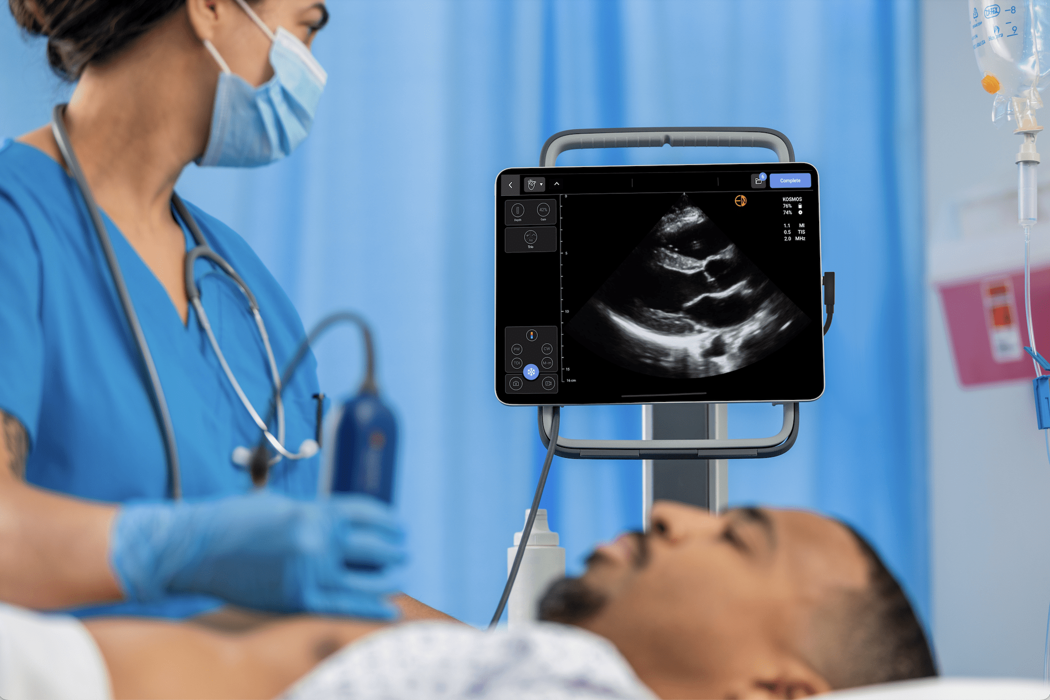
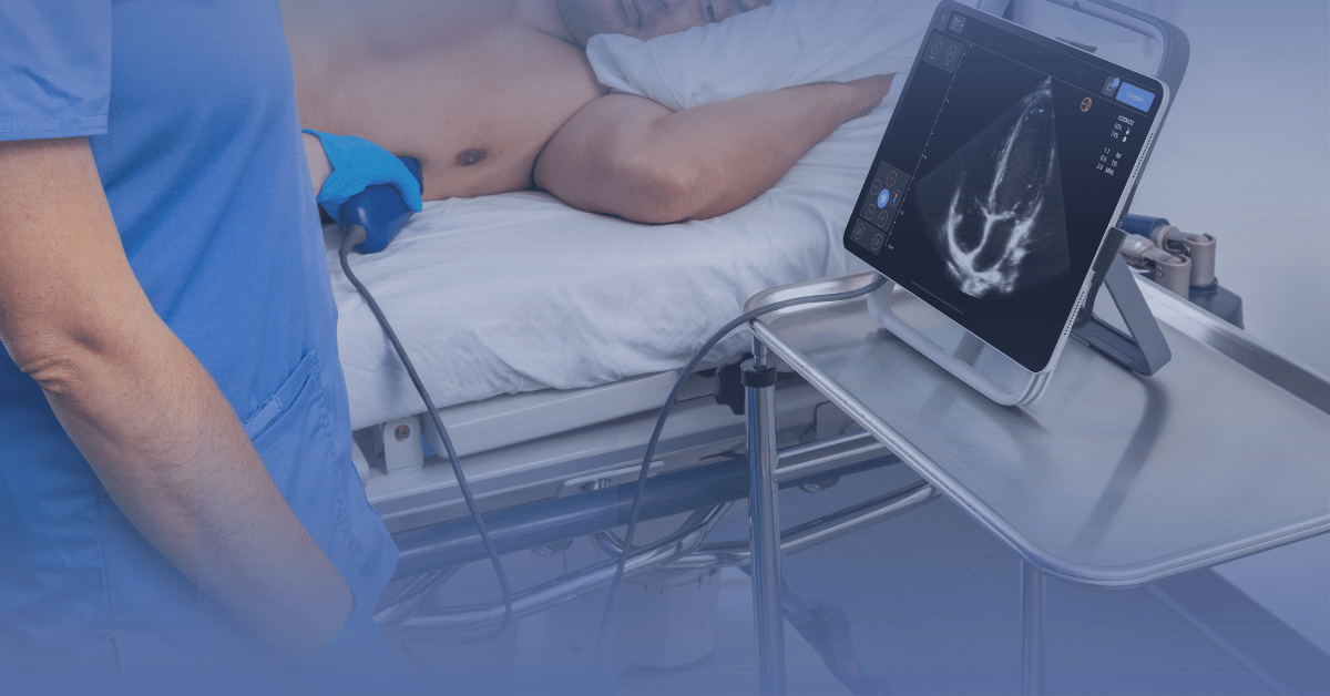
Blunt chest trauma can lead to a rare but life-threatening complication known as cardiac tamponade, particularly after high-contact sports such as football or cycling. Prompt identification and treatment are essential, and point-of-care Ultrasound (POCUS) is a valuable interoperable tool for early detection. The main sonographic signs of cardiac tamponade include pericardial effusion, right atrial or ventricular diastolic collapse, and inferior vena cava (IVC) dilatation, all of which can be assessed within seconds with conventional imaging windows (1).
The subxiphoid and parasternal long-axis views are beneficial when visualizing effusions and assessing cardiac chamber dynamics in the trauma setting. Doing what POCUS does best, dynamic to image guidance, immediate differentiation of cardiac tamponade from other causes of shock is made, and a timely intervention, such as pericardiocentesis, can be guided. POCUS has been further highlighted as an essential tool for cardiac tamponade diagnosis in emergency and critical care studies, demonstrating its relevance beyond the field.
Other Thoracic Uses
Outside of cardiac evaluation, POCUS is uniquely suited to the diagnosis of pneumothorax, a frequent complication of chest trauma. Lung sliding is absent, and a characteristic sonographic sign of the barcode sign (on M-mode) is present. POCUS can also help identify rib fractures not seen on initial radiographs or differentiate them from muscular strains.
In cases of trauma-related vascular complications, Doppler ultrasound and compression ultrasonography may also be used to evaluate surrounding soft tissue for associated pseudoaneurysm or hematoma formation, further extending the diagnostic range of thoracic POCUS.
Fracture Diagnosis and Interventional Guidance
Fracture Imaging with Ultrasound
POCUS is gaining recognition and value as a diagnostic tool in fracture detection, particularly when traditional radiographs fail to yield definitive results. It allows direct inspection of cortical disruption, periosteal elevation, and overlying hematomas, which are the hallmarks of bone injury. This is especially useful in children and individuals who partake in sports, where stress injury can be missed with initial X-rays, as they may not readily display subtle or non-displaced pathology (1).
POCUS’s follow-up benefits include assessing callus formation in healing fractures, allowing for continued monitoring of the healing process without further exposure to ionizing radiation. Its portability and dynamic imaging abilities also lend themselves to sideline or clinic-based evaluations.
US-Guided Procedures in Family Medicine
POCUS also improves the safety and accuracy of procedures performed in the outpatient setting. Family physicians are increasingly applying ultrasound guidance to procedures such as hematoma aspiration, platelet-rich plasma (PRP) injections, and guided delivery of corticosteroids, particularly for musculoskeletal conditions. Real-time imaging enables clinicians to visualize needle placement, avoid vascular structures, and confirm treatment delivery at the target site.
Such procedures are guided by doppler ultrasound, which serves a vital function in detecting adjacent vessels and avoiding iatrogenic damage. In treating tissues that may be located adjacent to a rich vascular bed, consideration should be given to the risk of causing a pseudoaneurysm, especially in trauma or post-injury tissues. This underscores the utility of ultrasound as both a diagnostic and a procedural safety tool in family medicine (1).
Practical Considerations for Family Medicine Practice
Integrating POCUS into Primary Care Settings
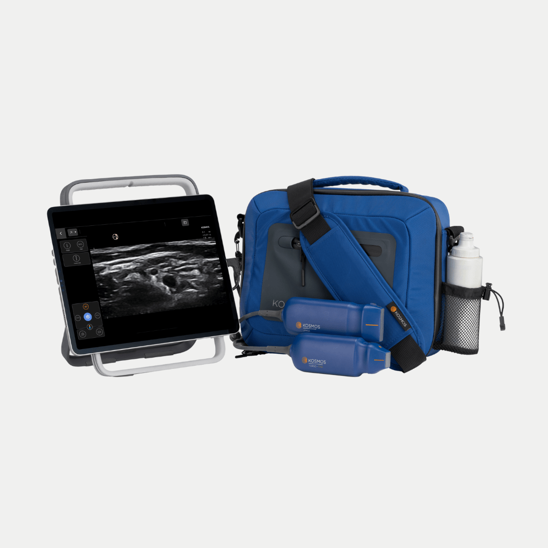
Integrating Point-of-care Ultrasound (POCUS) into family medicine practice has clinical and logistical benefits. Especially with contemporary point-of-care ultrasound (POCUS) devices that are small and ultramobile, ultrasound can even be used in various settings ranging from outpatient clinics and athletic sidelines to training rooms. This permutation allows expedient triaging and immediate diagnostic discourses and reduces the utilization of expensive imaging modalities such as MRI (1).
POCUS facilitates timely diagnosis and monitoring of various conditions, optimizing acute and longitudinal management with near real-time examination results from a cost and care-efficiency standpoint. However, to implement these successfully, structured training policies and standardized documentation procedures are essential for quality and consistency across users and the care environments.
Family physicians can use POCUS to assess life-threatening emergencies, including cardiac tamponade and vascular pathology like pseudoaneurysm, and it expands the physician’s armamentarium by allowing for compression ultrasonography for deep vein thrombosis in primary care. This positions POCUS as a strong tool for specialty providers and clinicians with general practices looking to broaden their horizons for diagnostic and procedural capabilities while maintaining a patient-centred approach (1).
Conclusion
The inclusion of point-of-care ultrasound (POCUS) for sports injuries provides family medicine providers with an immediate and accurate, yet affordable, opportunity for diagnosis. By guiding the assessment of muscle tears and ligament injuries, interpreting vascular complications like pseudoaneurysms, and detecting potentially fatal conditions like cardiac tamponade, POCUS improves clinical decision-making in diverse scenarios.
Doppler ultrasound and compression ultrasonography are among the technologies that enhance their use and provide accuracy in emergency and outpatient settings. As POCUS becomes more widely adopted, its incorporation into routine management will be necessary to address injuries promptly and effectively.
Frequently Asked Questions
1: How accurate is POCUS compared to MRI for sports injuries?
The diagnostic accuracy of POCUS has been high, especially for soft tissue and ligament injuries. That said, it has sensitivity and specificity similar to MRI when evaluating for ACL and PCL injuries. Although MRI remains the gold standard in many situations, POCUS is a trustworthy and fast first-line approach in almost all acute and follow-up evaluations (4,5).
2: Can Family Physicians use POCUS to identify vascular complications like pseudoaneurysm?
Yes, POCUS-trained family physicians can correctly diagnose pseudoaneurysms using doppler to note turbulent flow. These vascular complications must be diagnosed early to prevent morbidity and mortality, and thus, POCUS becomes an important asset not only in trauma care but also in post-intervention follow-up and primary care settings.
3: When should compression ultrasonography be used in sports medicine?
Compression ultrasonography helps screen for deep vein thrombosis (DVT) in immobilized or post-surgical athletes. It enables rapid diagnosis in a non-invasive, bedside manner that facilitates timely treatment, minimizing the risk of severe complications, including pulmonary embolism.
References
1. Lerchbaumer, M. H. et al, 2024. Ultrasound in sports traumatology. RoFo: Fortschritte Auf Dem Gebiete Der Rontgenstrahlen Und Der Nuklearmedizin, 196(5), 440–449.
2. Cassels, M., 2019. Point-of-Care Ultrasound as a Component of Preparticipation Screening of Athletes: A Systematic Review. Journal of Ultrasound in Medicine: Official Journal of the American Institute of Ultrasound in Medicine, 38(12), 3123–3130.
3. Draghi, F., et al. (2013). Muscle injuries: ultrasound evaluation in the acute phase. Journal of Ultrasound, 16(4), 209–214.
4. Ghosh, N., et al. (2017). Comparing point-of-care ultrasound (POCUS) to MRI for the diagnosis of medial compartment knee injuries. Journal of Medical Ultrasound, 25(3), 167–172.
5. Ahmadi, O., et al. (2024). The predictive value of point-of-care ultrasonography versus magnetic resonance imaging in assessing medial meniscal tears in patients with acute knee injury. Clinical and Experimental Emergency Medicine, 11(2), 188–194.
6. Hodgson, R. J., et al. (2012). Tendon and ligament imaging. British Journal of Radiology, 85(1016), 1157–1172.