Dr. Andre Kumar

Clinical Associate Professor, MD, MEd, Stanford Division of Hospital Medicine
How AI is Transforming POCUS: Exploring the Research of Dr. Andre Kumar
We recently sat down with Dr. Andre Kumar, Clinical Associate Professor at Stanford University who specializes in researching POCUS and AI with a focus on enhancing medical practices.
Dr. Kumar believes AI-assisted POCUS has the potential to revolutionize patient care, improve medical student training, and enhance physicians’ diagnostic capabilities.
Meet Dr. Kumar
Dr. Kumar, a Clinical Associate Professor at Stanford University, manages the Medicine Procedure and Consult Services, leads the Rathmann Fellowship in Medical Education, and co-directs the Advanced Clinical Skills course at Stanford University Hospital. He also co-founded the SHAPE Program, dedicated to enhancing hospitalist skills to meet modern healthcare demands.
In addition to these responsibilities, Dr. Kumar is a researcher specializing in POCUS and AI, with a focus on enhancing medical practices. He earned his MD from Tulane University, completed residency and chief residency in internal medicine at Stanford University, and holds a master’s degree in education (MEd).
Dr. Kumar has held several leadership positions during his career in medicine, including president of the Society of Hospital Medicine (SHM) Bay Area and director of the Stanford Internal Medicine Procedure Service. His leadership has been crucial to advancing medical education, research, and patient care.
Dr. Kumar’s journey into AI-assisted POCUS began during his internship, when he encountered a POCUS device brought by one of his attending physicians. Witnessing the real-time information and diagnostic capabilities from the device, he recognized its potential to revolutionize medical practice. This experience led him to specialize in POCUS and even study its effectiveness.
“It just completely opened up my eyes to what the profession can be, what we can actually do for our patients,” he said.
Putting POCUS to the Test: A Research Summary
In collaboration with colleagues from Stanford University School of Medicine, Dr. Kumar recently concluded his third randomized trial on Point-of-Care Ultrasound (POCUS) and its impact on patient care.
The study, titled “Acquisition of Cardiac Point-of-Care Ultrasound Images with Deep Learning: A Randomized Trial for Educational Outcomes,” sought to assess whether AI-enabled POCUS devices improve the competency of medical trainees with limited POCUS experience in acquiring apical 4-chamber (A4C) echocardiogram images.
The secondary outcomes of the study were to assess the quality of A4C images acquired using AI-enabled POCUS devices compared to non-AI devices, to evaluate the correct identification of pathology in A4C images with AI assistance versus without, and to measure the attitudes and confidence levels of participants in using POCUS devices with AI features.
Dr. Kumar and his colleagues conducted their study at a single academic institution from 2021-2022. Researchers randomized 43 internal medicine trainees with limited POCUS experience during an inpatient rotation to receive a POCUS device for two weeks. The device was either equipped with AI functionality (EchoNous, N=22) or without AI functionality (Butterfly, N=21).
The AI device provided automatic labeling of cardiac structures, guidance for optimal probe placement, and ejection fraction estimation. The primary outcome was the time to acquire an apical 4-chamber (A4C) image, and secondary outcomes included A4C image quality, correct pathology identification, and participant attitudes. They measured the data at baseline and two-week follow-up.
Key Findings and Future Implications
The study aimed to determine the effectiveness of an AI-powered device in improving the acquisition of A4C images. The results showed that the AI group outperformed the non-AI group in several key areas:
- Efficiency in Scan Acquisition: The study revealed that AI-powered devices significantly reduced the time that users required to acquire A4C images during follow-up assessments. This improved efficiency is pivotal for timely and accurate diagnosis in clinical practice, enhancing patient care by enabling quicker interventions.
- Enhanced Image Quality: AI-equipped devices consistently produced higher-quality A4C images, a crucial factor in precise diagnosis and treatment planning. Improved image quality may reduce the need for additional tests or procedures, streamlining patient care and potentially lowering healthcare costs.
- Improved Pathology Identification: Users of AI-enhanced devices demonstrated a higher accuracy rate in identifying specific pathologies, particularly reduced systolic function. This finding underscores the potential of AI to assist healthcare professionals in making more informed clinical decisions based on image analysis.
- Device Usage: Participants using AI devices reported more frequent usage, suggesting that AI can encourage active engagement with ultrasound technology. This increased utilization may contribute to enhanced proficiency among users over time.
- Participant Attitudes: Interestingly, despite the improved performance, both AI and non-AI groups reported similar attitudes towards the devices, with no significant differences noted. This highlights the importance of addressing trust and confidence issues in AI technologies through comprehensive training and education.
By harnessing artificial intelligence (AI) capabilities in point-of-care ultrasound (POCUS), this research offers a glimpse into the transformative potential of technology in healthcare. One of the most notable implications is the prospect of significantly enhancing healthcare professionals’ competency in acquiring and interpreting medical images.
The Big Idea: AI’s Role in Diagnosis and Training
As the demand for POCUS continues to grow, AI can serve as a powerful tool to bridge the gap between the training limitations of providers and the need for accurate and timely diagnosis. For example, AI-enabled POCUS has the potential to lessen the learning curve for new or inexperienced users, ensuring that healthcare practitioners of different experience levels can use advanced imaging techniques to improve patient outcomes.
Moreover, the study’s findings underscore the critical role that AI can play in increasing image acquisition efficiency, reducing scanning times, and improving image quality.
“You can see a potential where it can reduce delays for patient diagnosis,” Dr. Kumar said.
Furthermore, AI’s ability to help identify specific pathologies, such as reduced systolic function, highlights its potential as a valuable aid in clinical decision-making.
“One of the things that really drew me to point-of-care ultrasound was just seeing how we could quickly answer a lot of questions that we had about a patient,” Dr. Kumar noted.
However, through his research, Dr. Kumar has also found that the perception of AI is complicated. His study highlights the importance of addressing trust and confidence issues in AI systems. The research also emphasizes the need for comprehensive training and education to ensure healthcare professionals can fully harness the potential of these technologies.
Can AI-Enabled POCUS Help the Next Generation of Medical Students?
When Dr. Kumar and his colleagues train medical students using POCUS today, they often refer to the “feedback loop” that helps students learn effectively. This occurs when students are able to receive live guidance from an attending physician or senior colleague on their diagnostic technique.
But there are limitations on the feedback loop concept. For example, Dr. Kumar and his colleagues can’t personally oversee every resident and their usage of POCUS systems. This means students may be left without critical feedback on their technique at the moment of use.
“When I go off on my own and start scanning, that mentor may no longer be there, and I am, in many ways, scanning blindly or without substantial guidance and I lose that critical component of feedback for my own growth,” Dr. Kumar explained, citing the situation facing many students.
AI, however, can provide that instant feedback to the end user, and while it can’t replicate an instructor, it can close the gap in a busy or under-resourced clinical setting.
“Technology like this can have an enormous impact on healthcare by boosting expertise and helping to enhance the skills we need,” Dr. Kumar said. “And then you think about settings where there is no teacher, where someone really wants to use point of care ultrasound during a limited resource setting…that’s a particular area where you can really use this technology for their benefit.”
The Kosmos System that Dr. Kumar used in his research is a prime example of feedback enhancement. By seamlessly incorporating AI tools for real-time analysis and exam guidance, the Kosmos System echoes Dr. Kumar’s exploration of AI’s potential in aiding image acquisition and interpretation.
Additionally, the system’s inclusion of automated presets tailored for cardiology, abdominal, and lung applications simplifies the scanning process. This aligns well with Dr. Kumar’s emphasis on reducing the learning curve. Notably, the Torso-One Probe, equipped with phased array technology for cardiac and abdominal scanning, exemplifies how AI-assisted devices can enhance traditional teaching methods, offering superior guidance for probe placement and facilitating rapid pathology identification—both key aspects of Dr. Kumar’s research objectives.
Long-Term Vision for AI and POCUS in Healthcare
Dr. Kumar envisions a transformative role for AI in healthcare that promises to elevate diagnostic precision and operational efficiency. He underscores the importance of medical educators incorporating AI and POCUS training into medical curricula, recognizing the potential to significantly enhance patient care and healthcare delivery quality.
As a clinical educator, Dr. Kumar is acutely aware of the limitations of traditional medical education, especially concerning physical examination. He firmly believes that integrating POCUS and AI into medical education can provide medical students and residents with a more accurate and efficient means of diagnosing and treating patients. He advocates for POCUS devices to become a standard part of the physical examination curriculum as a way to enable doctors to enhance diagnostic accuracy and treatment decisions.
Dr. Kumar sees AI-assisted POCUS as a powerful tool for elevating patient care in medical practice. AI can help physicians interpret POCUS images more accurately and efficiently, facilitating faster and more reliable diagnoses. Additionally, AI can support doctors in tracking patient progress and predicting outcomes, enabling personalized treatment plans. There may even be applications for POCUS among non-medical professionals.
“Eventually, where I would really like to see the field go…is you put this technology in the hands of, let’s say, paramedical or even non-medical individuals and can augment their skills to a point where they can help increase the accessibility of care,” Dr. Kumar noted.
For example, he envisions a future where home health aides or nurses can use POCUS technology to provide remote scanning capabilities for cardiologists who may be unable to visit rural or isolated patients.
As a seasoned expert in POCUS and clinical medicine, Dr. Kumar deeply appreciates how AI can enhance the interpretation of ultrasound imaging, providing valuable insights for healthcare professionals. He firmly believes that integrating AI-assisted POCUS into medical education will empower future physicians to harness this technology to its fullest extent.
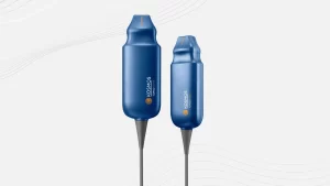
About The Kosmos Ultraportable Ultrasound System
The Kosmos Ultraportable Ultrasound System is a notable advancement in point-of-care ultrasound (POCUS) technology. It stands out for its compact form, high-quality imaging capabilities, and integration of advanced AI tools.
Unlike traditional ultraportable POCUS devices, the Kosmos System refuses to compromise on image quality. This system leverages cutting-edge CPU technology, proprietary ASICS, and high-performance PZT transducers to deliver exceptional image quality and durability.
One of its distinguishing features is the Torso-One Probe, which utilizes phased array technology. This probe enables cardiac and abdominal scanning using a single device, offering convenience and efficiency. Additionally, the system encompasses 2D, PW, CW, Color, and TDI features and access to various AI applications.
The Kosmos System also includes the Lexsa Probe, featuring a linear array suitable for small parts, lung, and vascular applications. This probe extends the system’s versatility with a 38mm aperture, 64/128 channels, and multiple imaging capabilities.
The Kosmos Bridge provides a high-quality display with intuitive scanning controls to facilitate ease of use. It allows users to capture, save, and export high-quality exams effortlessly. The system is HIPAA compliant, ensuring secure data handling.
Kosmos System compatibility with Android and iOS tablets further extends its utility. Users can experience high-quality imaging and harness deep learning AI capabilities on select tablets, making it adaptable to various clinical environments.
Reference:
- Baum, E, Tandel MD, Ren, C, Weng Y, Pascucci, M, Kugler J, Cardoza K, Kumar A. Use of Artificial Intelligence for Acquisition of Limited Echocardiograms: A Randomized Controlled Trial for Educational Outcomes. medRxiv. Preprint posted online on April 15, 2023. Doi:10.1101/2023.04.12.23288497