Going Beyond the Surface and Unveiling the Impact of Ultrasound on Diagnosis: A Conversation with Dr. Vimal Bhardwaj

Dr. Vimal Bhardwaj
MD
Luke Baldwin
VP of Global Marketing, EchoNous
Luke Baldwin, VP of Global Marketing, and our team at EchoNous recently had the opportunity to connect with Dr. Vimal Bharadwaj, a cardiac intensivist physician, researcher, and entrepreneur. Dr. Bharadwaj’s expertise in integrating ultrasound technology into patient care makes him a valuable voice in critical care medicine. In this discussion, Dr. Bharadwaj shares his expertise on the practical applications of point-of-care ultrasound (POCUS) in healthcare and discusses his recent research paper on Femoral Vein Doppler (FVD).
Dr. Bharadwaj’s journey illustrates the practical application of POCUS in healthcare. With over a decade of experience as a critical care physician, he emphasizes the importance of ultrasound in optimizing patient care, particularly in adult cardiac cases. His expertise in postoperative management and cardiac stabilization underscores the vital role of ultrasound technology in facilitating accurate diagnoses and informed treatment decisions. Leveraging ultrasound for precise medical interventions reflects a commitment to improving patient outcomes through evidence-based practice.
[Note: This interview has been edited for clarity and brevity.]
Luke Baldwin: It’s great to meet you again. For those who don’t know you, could you please introduce yourself and tell us about what you do and what you focus on?
Vimal Bharadwaj, MD: Sure, thank you. I’m Dr. Vimal Bharadwaj, a critical care physician with 12 years of clinical experience. I work at Narayana Health, specifically in the city of Narayana, which houses a 1400-bed facility, making it one of the world’s largest cardiac hospitals.
We handle a variety of procedures daily, including around 35 to 40 CAGs (Coronary Angiograms), valve replacement surgeries, heart transplants, and lung transplants. As a cardiac intensivist, I specialize in stabilization and postoperative care for transplant patients and manage Venoarterial (VA) Extracorporeal Membrane Oxygenation (ECMO) and Venovenous (VV) ECMO.
My training and practice are centered on adult cardiac care, while we have a separate pediatric division for complex heart surgeries. My particular interest lies in utilizing point-of-care ultrasound to diagnose various pathologies. I firmly believe in the importance of looking inside to treat patients accurately, and ultrasound provides an invaluable tool.
Luke Baldwin: So, tell me a little about your first encounter with ultrasound. You’re obviously a huge advocate and point-of-care ultrasound user; what was your first exposure to ultrasound? How did you become so passionate about using it on every exam?
Vimal Bharadwaj, MD: So, whenever we see a sick patient, the clinical examination might not fully reflect what’s happening inside. When I started noticing patients coming back with issues like pulmonary edema or bloating, it became clear that clinical findings alone might not reveal the true internal picture. Ultrasound was starting to be introduced into the ICU when I began my specialization. As I delved deeper into its nuances, I realized its immense value.
With proficiency in ultrasound, diagnosing life-threatening pathologies became a matter of minutes, sometimes even seconds. This capability boosts confidence and enables timely and life-saving clinical decisions. I’ve encountered cases where we instantly diagnosed conditions like tension pneumothorax and saved lives.
Ultrasound has become an indispensable tool for me. It enhances my understanding of dynamic organ pathologies and guides critical treatment decisions, such as adjusting ventilator settings or assessing the effectiveness of cardiac support. Witnessing how ultrasound-informed clinical judgments directly impact patient outcomes is truly exciting.
Luke Baldwin: Just circling back to your training in ultrasound, it seems you’re scanning some complex pathologies. Can you walk us through your training process? Did it happen during medical school or did you learn on-the-job?
Vimal Bharadwaj, MD: In India, we specialize in critical care as part of our advanced training. I did my anesthesia specialty training in an institute where ultrasound was just introduced. It was in an institute where ultrasound was not widely available in other parts of India. We began using ultrasound for nerve blocks.
During my critical care specialization, ultrasound became part of our curriculum. We had to undergo a structured ultrasound training program with various protocols, like the blue lung protocol. We had to perform at least 10 or 15 scans in front of a consultant, who would monitor us during the procedure. The consultant had to countersign our logbook, and ultrasound questions were part of our examinations.
We underwent rigorous ultrasound training, and now I’m a faculty member for critical care echocardiography, which was recently launched in India. Training is a continuous process, and we’ve been fortunate to have structured ultrasound training programs.
Luke Baldwin: Very good. I’d love to hear about your research on Femoral Vein Doppler and its implications for assessing right heart function. What prompted you to explore this area?
Vimal Bharadwaj, MD: As a cardiac intensivist, right ventricular failure is one of the key factors determining postoperative mortality. Whenever a patient comes in post-cardiac surgery, windowing for ultrasound isn’t always perfect, and right ventricular failure is quite prevalent. I came across a chapter on venous congestion by Dr. Philippe Rola and his team from Montreal, which intrigued me. Despite veins housing 70% of blood volume, there’s been more focus on arterial side issues. This raised questions about why venous issues aren’t part of our daily practice.
I connected with Dr. Philippe Rola about his work on venous congestion scoring (VExUS), which was being proposed. I conducted India’s first research on VExUS in cardiac failure patients. It was challenging; VExUS training took a month. Seeking alternatives, I found an article from 1922 by Dr. Kerr correlating venous pulsations with right heart failure. Further, recent case reports suggested a correlation between VExUS and femoral venous pulsations.
My research led to a prospective study comparing VExUS with Femoral Venous Doppler and central venous pressure (CVP) in post-cardiac surgical patients. Femoral Vein Doppler was more sensitive than VExUS in assessing venous congestion, correlating 86% with Doppler. We published our findings in an ultrasound journal, and I now routinely perform Femoral Venous Doppler.
We’re researching whether Femoral Venous Doppler correlates with right ventricular-pulmonary artery coupling. Preliminary results suggest a correlation, offering insights into right heart function without echocardiography.
Another ongoing study involves dialysis patients, assessing whether normalization of Femoral Venous Doppler improves quality of life. Additionally, we’re comparing Femoral Venous Doppler with VExUS in acute physiology and chronic health evaluation (APACHE) scores.
“Femoral vein pulsatility: a simple tool for venous congestion assessment” provides critical information for inotropic support decisions, fluid management, and right heart function assessment, all within a minute. Its simplicity makes it accessible for training new ultrasonographers.
We’re also planning studies on inter-observer variability and the learning curve between Femoral Venous Doppler and VExUS. Within the next few months, we’ll have more articles on the utility of Femoral Venous Doppler.
Luke Baldwin: I am familiar with VExUS grading, and I am sure most of our audience is as well, but I’m curious about Femoral Venous Doppler.
Let’s say you begin with Femoral Venous Doppler. What’s the next step? If you observe pulsatility, do you continue with the VExUS protocol, examining the hepatic or portal vein? Or do you typically stop at FVD? What’s your usual approach after noticing a positive FVD?
Vimal Bharadwaj, MD: Yeah. So, any ultrasound examination of a single entity is just a piece of the puzzle. It has to be correlated. I invariably perform VExUS, and I’ve been nicknamed “VExUS” because of how often I use it. My examination is only complete after considering the clinical profile, Femoral Venous Doppler, and right heart function.
It’s never a complete examination by looking at one aspect; continuity is key. During the initial examination, I assess the patient’s clinical profile, right and left heart function, and perfusion parameters. If needed, I start inotropic agents and diuresis, followed by serial Femoral Venous Doppler and arterial blood gas analysis.
Once pulsations disappear, I can confidently adjust inotropic support. The first examination should always provide a complete patient profile; it’s just one piece of the puzzle, not the entire answer.
Luke Baldwin: Got it. Based on your research and experience, how should those familiar with VExUS incorporate Femoral Venous Doppler into patient management?
Vimal Bharadwaj, MD: To authenticate Femoral Venous Doppler, you must practice on five to six cases to gain proficiency. You’ll understand what constitutes abnormality once you understand venous flow and pulsatility. With more practice, Femoral Vein Doppler becomes more straightforward, especially in post-cardiac surgical settings where ultrasound windows may not be optimal.
If central venous access isn’t feasible, Femoral Vein Doppler is always an option. We’ve discussed incorporating a Femoral Vein Doppler score into VExUS to enhance objectivity in diagnosing venous congestion. More parameters improve diagnostic accuracy, reducing ambiguity.
One flaw with VExUS is its reliance on an absolute IVC size of 2 cm, which may not always apply. If the clinical profile suggests venous congestion and the IVC is non-collapsing, but smaller than 2 cm, Femoral Vein Doppler provides valuable information.
Further research is needed to define venous congestion objectively, but Femoral Vein Doppler and VExUS offer insights into venous condition.
Luke Baldwin: You mentioned it’s easy to learn, and everyone falls along a spectrum in their experience with ultrasound. Do you see an opportunity for novices to begin with Femoral Venous Doppler? Could this be a starting point or something they learn early to help monitor patients? Or do you think it’s still for expert users?
Vimal Bharadwaj, MD: I must reveal something to answer this question. I’m conducting a multicenter trial in India on VExUS-guided diuretic therapy to reduce hospital readmissions in heart failure patients. The sample size is 500 patients across four hospitals.
Initially, convincing cardiologists to use VExUS for therapy optimization was a significant challenge. However, acceptance has grown as we’ve enrolled 80 patients and demonstrated its utility.
So, any newer thing is challenging for people to accept. Everyone in our institute is getting comfortable with VExUS because we have already enrolled 80 patients. I suppose the same thing was good for me as well.
As you can see, the utility of Femoral Venous Doppler is quite enormous if you think beyond the text. What if we show a femoral vein? Because it is superficial, it can have many implications for building an artificial intelligence algorithm, whether it be a feedback mechanism in dialysis patients or those undergoing dialysis; we do not know how much fluid to remove. What if we have an ultrasound and the femoral vein and a feedback mechanism between the ultrasound and the dialysis machine, and once it detects the pulsations coming down, that’s when it can stop ultrafiltration?
There are tremendous opportunities here because it is superficial and easy to detect so you can get many applications out of this femoral vein. This is just one application, not only in heart failure patients for titration of heart failure treatment, so these are also other added applications that can be explored in the future.
It takes 5 to 10 cases under supervision. When I was teaching my residents, I wanted to see how good they were, so I told them to show their Femoral Vein Doppler findings. I could see that almost all of them were very comfortable with VExUS, but some, especially novices, had difficulty visualizing the IVC properly. For experienced users, it should be easy, but for novices, it poses some challenges.
The human mind always takes an easier path; if you cannot get it, you might not pursue it, but in 10 out of 10 cases, you should find some signal in the Femoral Vein Doppler. I do not see why you would not be able to visualize it unless the patient is morbidly obese, a hematoma or some other condition is obscuring it, or there is a DVT or some other issue.
But there is no other good reason you should be unable to visualize it. So, in at least 9 out of 10 cases, it’s very easy to visualize something, and once it is easier, the acceptability rate also goes up. That’s what I tremendously believe. Once we propagate this concept, who knows what more research, some groundbreaking or pathbreaking research work might entail, and you might find a phenomenal understanding of the right heart and venous condition.
Luke Baldwin: I was curious to hear you mentioned the limitations of FVD. Can you expound on that? What are some of the limitations that you’ve observed so far?
Vimal Bharadwaj, MD: The femoral vein cannot be used in tachypneic patients because you need a supine patient that mirrors the IVC. The factors that affect the IVC should also affect the femoral vein. Abdominal pressure can influence the IVC, not only from the weak pressure but also from the thoracic complaints. The same factors should affect FVD as well.
So, it cannot be used in patients with low thoracic complaints, those who require high PEEP on ventilators, and those who are quite technical using accessory respiratory muscles. These patients have quite an active diaphragm, so the diaphragm is just falsely compressing the IVC, and you will find the same finding over there.
So, all the exclusion criteria for VExUS fit apart from deep vein thrombosis. Yes, common iliac vein thrombosis. Also, you might find a Femoral Vein Doppler is not sending the signals of venous condition and sometimes the left femoral vein. [In that case,] you cannot use it; it has to be the right common central vein because it is in direct continuity with the IVC. There is only a single valve between the IVC and the right common femoral vein, which becomes incompetent whenever there is a venous condition.
It reflects the changes in IVC quite accurately compared to the left common femoral vein. So, these are some exclusions or considerations one should make when screening for venous conditions using formal Venous Doppler. None of the ultrasound examinations of a single parameter is accurate enough, 100% sensitive, or specific. That’s why more and more venous examinations are recommended to ascertain venous conditions.
Luke Baldwin: Right. To get a clearer picture, it’s like a single piece in the puzzle when you look at the broader scope.
Vimal Bharadwaj, MD: Yes, cirrhotic patients and those with intraabdominal hypertension might present challenges. In such cases, we might observe a falsely collapsing common femoral vein due to volume congestion and active diaphragm involvement.
Luke Baldwin: Tell me more about the ultrasound capabilities required for this exam. What specific features do you look for in the ultrasound equipment?
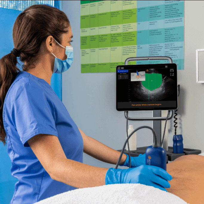
Vimal Bharadwaj, MD: Yeah, basically, we need reliable venous Doppler, so we are looking for a linear probe with good spectral Doppler, preferably a cardiac probe. I prefer a mobile ultrasound setup that is easy to carry and has both the vascular and cardiac probes on either side. So, that should suffice for 90% of my patients. I rarely use an abdominal probe.
However, with cardiac and vascular probes, I can look at the heart and veins with the linear probe, which is my basic requirement. Especially for the VExUS multicenter trial, since we have 500 patients to screen, carrying a bulky machine everywhere is quite strenuous. Doing this screening in the wards is also laborious, so having a mobile ultrasound will be quite an advantage.
Eventually, you would look at titrating in heart failure patients who come on an outpatient clinic basis. Just one essential examination, and you can be certain whether you have fine-tuned their heart failure treatment to better success. So, a portable ultrasound will be quite useful.
Luke Baldwin: Yeah, portability is essential, and obviously, spectral Doppler is important. When interrogating the heart, are you also using CW Doppler at that point?
Vimal Bharadwaj, MD: With the right heart, I do a thorough assessment, not only of ataxia but also of the fractional area change. I look at the septum along with peer pressure estimation using continuous wave (CW) Doppler. With right heart failure, you can get falsely low peer pressure, so I also look at the pulmonary artery. So, both pulse wave and continuous wave Doppler will be quite desirable.
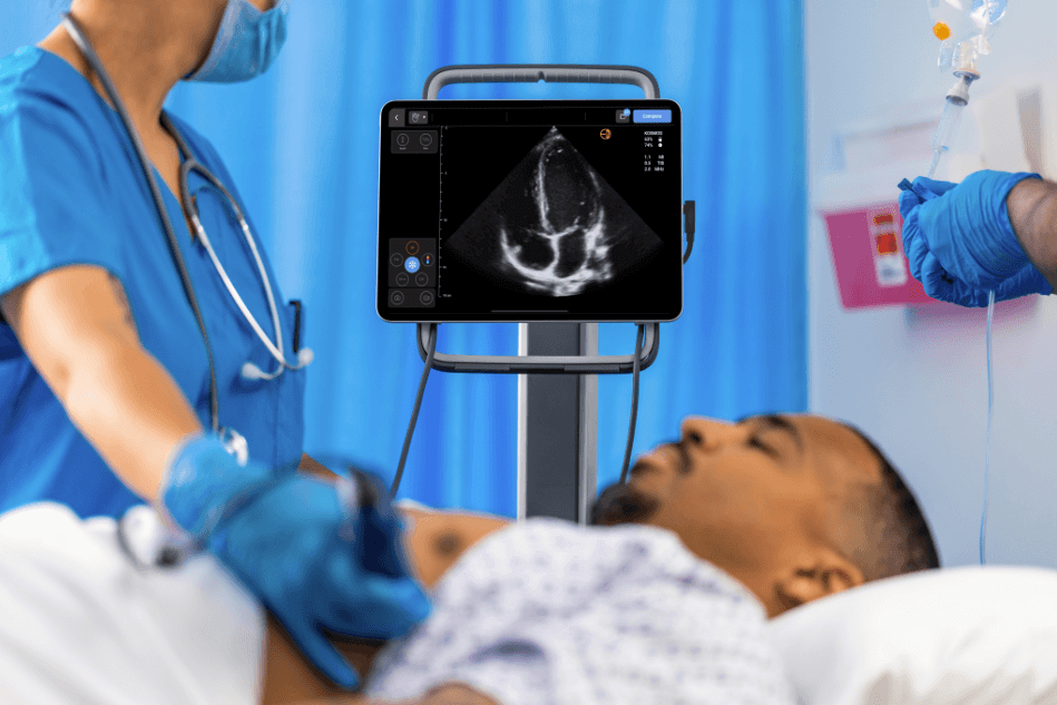
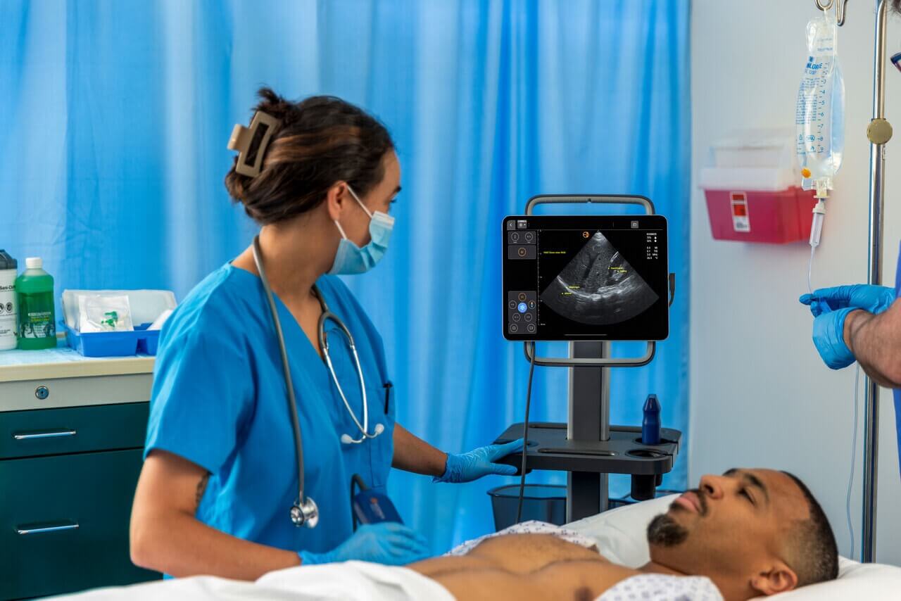
Luke Baldwin: One of the key goals in designing Kosmos was to ensure it was a comprehensive system. Many portable systems offer only partial solutions, leaving gaps in terms of what questions you can answer. With Kosmos, we aimed to provide a complete solution by including CW Doppler, PW Doppler, TDI, and other measurement features and calculations. So, if you had an issue, you wouldn’t need to pause the exam, and go find a full cart-based system to finish the exam; you could keep going with Kosmos all at once. It’s good to hear that we’ve got the right mix of capabilities, so you won’t be limited at the bedside.
Vimal Bharadwaj, MD: TDI is quite valuable in routine cardiology assessments. It provides essential indices for evaluating systolic and diastolic function, particularly for assessing the right heart. Combining TDI with other tools like echocardiography offers a comprehensive approach to cardiac evaluation.
Luke Baldwin: Excellent. Considering the future, could you elaborate on the implications of your research? You’ve mentioned ongoing studies. What further impact do you foresee this research having on patient management?
Vimal Bharadwaj, MD: Diuretic management plays a crucial role in patient care, as highlighted in the ACC guidelines. Inappropriate or inadequate diuretic therapy often leads to hospital readmissions. Using ultrasound, we can precisely manage diuretics by evaluating parameters of illness condition, thus improving patients’ quality of life.
Accurate diagnosis of conditions like right heart failure, particularly in post-cardiac surgical patients, is vital. Additionally, research on Doppler and sepsis offers insights into volume overload states commonly seen in septic shock patients. This research aids in timely diagnosis and resuscitation.
Addressing the emerging topic of de-resuscitation is significant. Establishing precise endpoints for de-resuscitation can greatly benefit patient outcomes. We can develop protocols and even artificial intelligence algorithms to predict patients by integrating physiological correlations.
This approach considers parameters like ATI percentage, renal system index, and volume condition to determine the need for interventions like dialysis. The implications of this research are vast, and we anticipate significant advancements in the field in the next few years.
Luke Baldwin: Thank you. You mentioned Dr. Philippe Rola and the global community of sharing knowledge. What is the best way to spread the word or advice for researchers to get their work noticed globally?
Vimal Bharadwaj, MD: The Montreal Heart Institute, where Dr. Rola and I have spoken, provides a platform for sharing valuable research. However, for research to truly make an impact, it needs to reach a wider audience. This involves publishing multiple papers in reputable journals and leveraging social media platforms for dissemination. Social media plays a significant role in amplifying research visibility. Once a concept gains traction, as we’ve seen with VExUS and hemoglobin, it becomes widely accepted and integrated into training curriculums, as observed in ultrasound workshops in India. Collaboration on an international scale also fosters the exchange of ideas and opens avenues for future research endeavors.
Luke Baldwin: We appreciate your time and are certainly big fans of your work. We’ll continue following you as well. If there’s an opportunity with some of your future research, if you want to do this again and talk about a new publication, the door’s always open. We’d be happy to have you.
Vimal Bharadwaj, MD: Excellent. I appreciate this opportunity for you to reach out and recognize the work. It means a lot. And thanks for talking to me; it felt so good to discuss the research.
To learn more about Kosmos, contact us today!