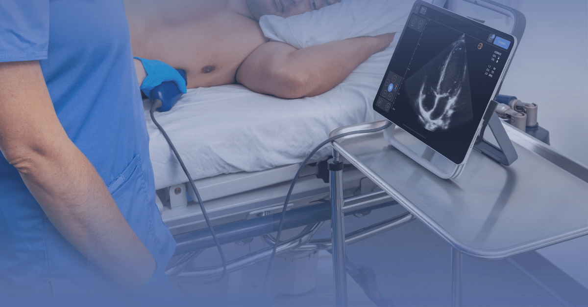
Could Ultrasound Be the Next Vital Sign?
Cardiac ultrasound aligns interestingly with modern cardiology: beyond its diagnostic features, it expands the boundary of full-spectrum cardiac assessment. With the introduction of point-of-care ultrasound (POCUS) modalities, cardiology is now enjoying a technological revolution.
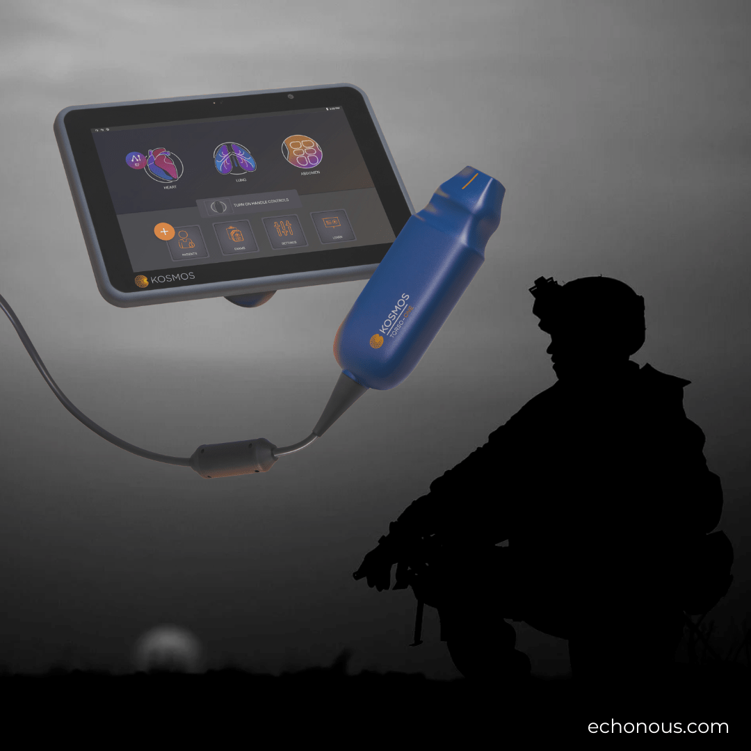
POCUS complements physical cardiac evaluation with less labor cost and more technical accuracy. Ultra-portable ultrasound devices operated by clinicians can enhance cardiac architecture imaging.
Available clinical studies supporting its integration into modern medicine have demonstrated incredible results on how POCUS can improve the detection of various cardiac anomalies with close to zero error rates.
Key Takeaways
- Point-of-care ultrasound (POCUS) is transforming cardiac care with portability and real-time imaging
- It is now being considered a new “vital sign” due to its ability to provide crucial insights into cardiac function
- POCUS excels in detecting heart conditions like valve abnormalities, cardiomyopathies and acute heart failure
- It enables rapid bedside assessments, reducing dependency on traditional imaging tools
- Technologies like color Doppler and real-time annotations elevate POCUS’s accuracy, enabling precise cardiac anatomy and function assessments
Table of contents
Clinical Evidence Supporting POCUS in Cardiac Care
Researchers have reported POCUS input in detecting left ventricular systolic dysfunction(1), pleural effusions(2), valvular abnormalities(3), pulmonary edema(4), and systemic venous congestion(5).
Compared with conventional cardiac imaging methods, POCUS offers quick, accurate results that might either complement a physical examination or, in many cases, aid diagnosis without physical findings.
In these diagnostics cases, ultrasound capabilities correctly identify tissues and cellular abnormalities, prompting noticeable variations in normal physiology. For instance, an enlarged left atrium suggests a possible elevated left heart filling pressure, a vertical B-line in the lung fields suggests pulmonary edema, global or regional wall motion abnormality suggests left ventricular systolic dysfunction and a dilated inferior vena cava suggests possible systemic venous congestion.
Today, advanced ultrasound capabilities can capture minor irregularities in cardiac function. Image enhancement features and color Doppler techniques are particularly essential in cardiac ultrasound.
Cardiac Ultrasound as a Vital Sign
With POCUS’ wide adoption and clinical positioning today, clinicians are ruminating on a controversial point:
‘Could point-of-care ultrasound be the next vital sign?’
Vital signs are the first clinical examinations required in patient triage; they inform the clinician of the degree of deviation from the positive baseline.
Temperature, blood pressure, respiratory rate, and pulse rate are the traditional vital signs.
In modern medicine, vital signs are not just metrics; the degree of vital signs abnormalities directly predicts patients, long-term care outcomes, symptoms severity, and emergency department re-admission probability(6).
Based on the clinical descriptions of ‘vital signs’, researchers argue that a few other parameters fit this description.
For instance, in many emergency departments in the United States, ‘pain’ is widely considered the fifth vital signal. Although subjective in reporting, pain severity reflects the patient’s comfort level and biologically informs on the severity of inflammation and tissue damage. Also, there are multiple clinical studies supporting pulse oximetry and smoking status as useful vital sign parameters – both significantly impact patient outcomes.
These studies and arguments prove a simple point: In clinical settings with multiple capabilities, ‘vital signs’ may capture and describe parameters that directly inform patients’ outcomes. On this premise, point-of-care ultrasound strategically belongs in the vital sign “Hall of Fame.” The wide adoption of POCUS in nephrology, cardiology, infectious diseases, intensive care, and emergency care signals a possible, gradual shift to advanced imaging capabilities in clinical assessments(7).
Vital Signs in Cardiology: POCUS Offerings
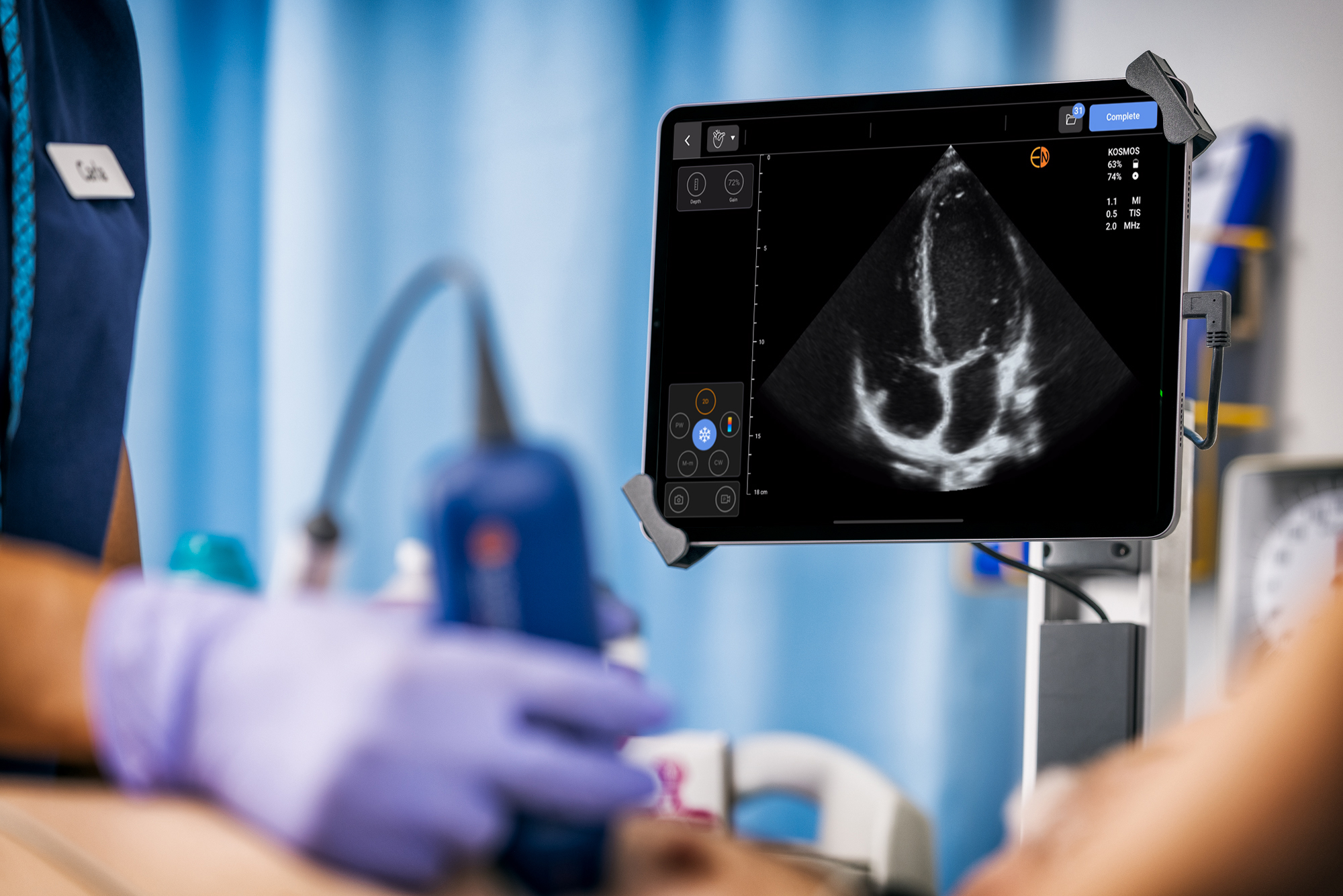
Cardiology thrives on accurate clinical assessments of cardiac functioning. POCUS modalities in cardiology leverage sound patterns to capture detailed images of the heart (and associated vessels) at different depths and pixel levels. Imaging outputs provide real-time insights into the heart’s anatomy and function.
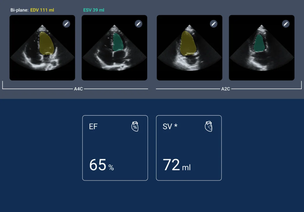
POCUS offerings extend beyond the images; they also offer enhanced views of the heart movement, providing information on valve function, wall motion, and ejection fraction.
These parameters are essential to patient management, guiding cardiologists on treatment decisions like the need for surgery or the need for medication withdrawal(8).
Some use cases support POCUS’s introduction as a vital sign in Cardiology. These include:
1. Diagnosing Valve Abnormalities
Advanced imaging features on POCUS machines capture non-static comprehensive images of the heart valves. Annotation features can also reveal thickening and calcification of the valve leaflets and changes in the directional movement of blood. Abnormal blood motion visualizations inform clinicians of possible regurgitation or valvular stenosis.
These spot checks help clinicians quantify the severity of blood motion disruptions and valvular defects. Essentially, information from cardiac ultrasound complements symptom assessment in making accurate diagnoses, evaluating the effectiveness of treatment regimens, and determining disease prognosis.
In a dynamically changing field of cardiovascular care, there are few limits to what POCUS could achieve for better patient outcomes.
2. Detecting Cardiomyopathies
Cardiomyopathies weaken the heart muscles, causing abnormalities in the heart’s blood-pumping functionality. Depending on severity, cardiomyopathies may also stiffen the heart, enlarging and thickening different sections. In worst-case scenarios, cardiomyopathies cause complications like stroke, cardiac arrest, cardiogenic shock, arrhythmias, and heart failure. Cardiomyopathies present a huge healthcare burden in cardiology.
POCUS’ capabilities in examining the cardiac musculature position it as an option for detecting cardiomyopathies. Compared to physical examination, POCUS offers better accuracy. It directly informs clinicians of how heart muscle changes affect function, structure, and size.
Combined with real-time annotation and information on blood flow, POCUS can help clinicians distinguish between the different forms of cardiomyopathies, including restrictive, ischemic, arrhythmogenic right ventricular dysplasia, left ventricular non-compaction, and dilated myopathies.
Post-diagnosis assessments are important in managing cardiomyopathies. Advanced POCUS capabilities also help quantify changes in the left ejection fraction and track wall thickness – these measures help assess disease progression. As a vital sign, cardiac ultrasound can detect subtle structural changes and improves the accuracy of detecting early signs of cardiomyopathy.
3. Screening Acute Heart Failures
Emergency presentations of acute heart failure may be clinically ambiguous in many cases. The differential diagnoses, depending on age group, lifestyle, medication, and family history are numerous. Despite all these variables, Acute Heart Failure requires rapid diagnosis and intervention.
Conventionally, clinicians rely on chest X-rays and physical examinations. Cardiac ultrasound may revolutionize the approach to diagnosing acute heart failure.
POCUS is portable, swift, and advanced. Its imaging capabilities also easily assess the common triggers of heart failure, including ventricular ejection dysfunctions, wall motion abnormalities, and valvular heart diseases.
It aids rapid diagnosis in emergency settings by helping clinicians understand hemodynamic status and pinpoint the underlying mechanism of heart failure. Its ability to detect pulmonary edema and visualize B-lines expedites treatment and improves survival rate. Bedside cardiac ultrasound allows for rapid and precise screening, granting cardiologists enough information to decide on the best treatment approach.
POCUS also helps cardiologists assess atrial size, analyze ventricular volume, and determine the dimensions of cardiac chambers. In the high-stakes clinical setting of cardiology, POCUS inclusion as a vital sign holds tremendous potential for optimizing patient outcomes.
Get Started: Include POCUS in Your Practice
Advanced point-of-care ultrasound systems like Kosmos provide cardiologists and other physicians with the ability to gather the clinical insights essential to optimal patient care.
Compared to conventional lab imaging, Kosmos’s AI-enabled workflows and advanced Doppler (including continuous wave) capabilities are designed to offer swift response as needed in a high-paced, intensive care setting like cardiology. Its potential offers a new world of possibilities.
If you are looking to get started with POCUS today, schedule a Kosmos demo! Our team of POCUS experts would love to learn more about your medical practice and how best to incorporate the right point-of-care ultrasound solution for you.
References
1. Motazedian, P., Marbach, J.A., Prosperi-Porta, G. et al. Diagnostic accuracy of point-of-care ultrasound with artificial intelligence-assisted assessment of left ventricular ejection fraction. npj Digit. Med. 6, 201 (2023). https://doi.org/10.1038/s41746-023-00945-1
2. Zaki, H. A., Albaroudi, B., Shaban, E. E., Shaban, A., Elgassim, M., Almarri, N. D., Basharat, K., & Azad, A. M. (2024). Advancement in pleura effusion diagnosis: a systematic review and meta-analysis of point-of-care ultrasound versus radiographic thoracic imaging. The ultrasound journal, 16(1), 3. https://doi.org/10.1186/s13089-023-00356-z
3. Hammadah, M., Ponce, C., Sorajja, P., Cavalcante, J. L., Garcia, S., & Gössl, M. (2020). Point-of-care ultrasound: Closing guideline gaps in screening for valvular heart disease. Clinical Cardiology, 43(12), 1368-1375. https://doi.org/10.1002/clc.23499
4. Kimura, B. J., & Nayak, K. R. (2020). “Asymptomatic” Flash Pulmonary Edema by Point-of-Care Ultrasound: A Novel Bedside Finding of Transient Global Ischemia. JACC. Case reports, 2(10), 1545–1549. https://doi.org/10.1016/j.jaccas.2020.06.029
5. Koratala A. (2022). Evaluation of Venous Congestion Using Beside Ultrasonography by the Nephrology Consultant: The VExUS Nexus. POCUS journal, 7(Kidney), 17–20. https://doi.org/10.24908/pocus.v7iKidney.15341
6. Sapra A, Malik A, Bhandari P. Vital Sign Assessment. [Updated 2023 May 1]. In: StatPearls [Internet]. Treasure Island (FL): StatPearls Publishing; 2024 Jan-. Available from: https://www.ncbi.nlm.nih.gov/books/NBK553213/
7. Alejandro Díez-Vidal, Patricia Martínez-Martín, Borja González-Muñoz, Yale Tung-Chen, Point-of-care Ultrasound in Infectious Diseases: Current Insights and Future Perspectives, Clinical Infectious Diseases, Volume 79, Issue 2, 15 August 2024, Pages 420–429, https://doi.org/10.1093/cid/ciae285
8. Herbst MK, Velasquez J, Adnan G, et al. Cardiac Ultrasound. [Updated 2022 Nov 21]. In: StatPearls [Internet]. Treasure Island (FL): StatPearls Publishing; 2024 Jan-. Available from: https://www.ncbi.nlm.nih.gov/books/NBK470584/
