POCUS Echocardiography at the Bedside for Hospitalists
Point-of-care ultrasound (POCUS) is rapidly becoming a go-to tool for hospitalists. It provides immediate assessment of patients and can significantly reduce the time it takes to reach a diagnosis, which in turn can lead to a number of efficiencies for the hospital.
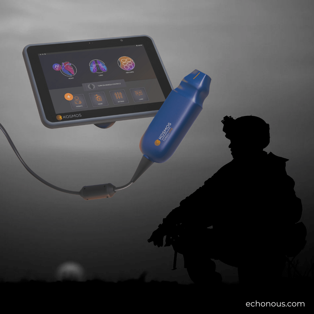
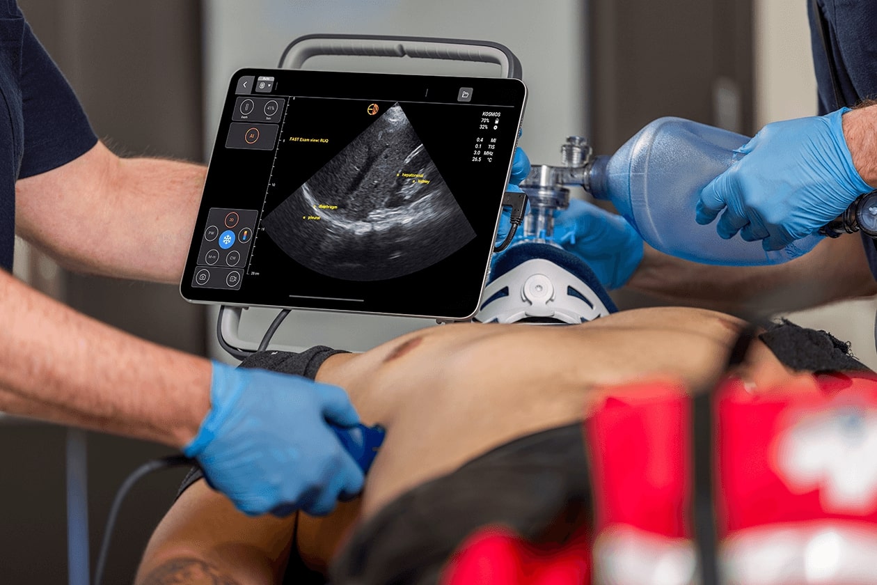
Compact, portable, AI-enabled POCUS solutions, like Kosmos, can be a game changer because these can empower clinicians to obtain real-time cardiac assessments, making medical decisions more informed and accurate. POCUS devices with echocardiography capabilities are a fraction of the cost of traditional ultrasound cart systems.
In clinical practice, a common thing clinicians may be looking for with a POCUS cardiac echo is to gauge left ventricular systolic function. And, with the right skills, and a little help from AI, the operator can assess most of the cardiac structures with a high level of accuracy and detail in just a few minutes.
Schedule your demo today and experience the future of patient care with Kosmos Plus
Table of contents
The Role of POCUS in Heart Failure
Due to the increasing number of patients with heart failure, and the shortage of cardiologists to treat them, the reality is that patients with heart failure undergo a formal ultrasound maybe only once a year (if that), or only when they are admitted to the hospital. Reviewing previous scans may not be the most illuminating when treating their current symptoms.
Treating these types of heart conditions based on vitals and physical exam alone can feel like driving through dense fog, especially when time with the patient is limited, they’re riding on the border of unstable, and the rounding cardiologist has 30+ more consults to go.
Portable echocardiography can play a crucial role in addressing and managing heart failure, as oftentimes waiting for a formal ultrasound can significantly delay care.
Hospitalists are finding incredible value in being able to perform bedside cardiac echos. These exams offer real-time insights, making diagnosis more efficient, treatment more direct, and prognosis much more accurate.
In heart failure, portable echocardiography can help in determining left ventricular ejection fraction (LVEF). LVEF is a key measurement based on which clinicians can classify patients into 3 groups:
- Reduced ejection fraction (HFrEF)
- Mildly reduced ejection fraction
- Preserved ejection fraction (HFpEF)
POCUS can help clinicians in identifying mitral regurgitation (MR), right ventricular dysfunction, and diastolic function as well, which are important prognostic indications, especially if the physician is scrambling to pick pressors during a resuscitation, or worry that the patient is falling into shock. For hospitalists that are new to ultrasound or worried about the learning curve, AI-assisted POCUS devices, like Kosmos, may help hospitalists to make more informed medical decisions, faster.
Interested in learning more about how to use POCUS in your clinical practice? We’ve got you covered.
POCUS to Monitor Pericardial Effusions
Cardiac tamponade is a medical emergency – something that can easily be avoided with regular sonograms, depending on the patient. It is characterized by fluid or blood accumulating in the pericardial sac, which leads to a decrease in cardiac output and eventual shock.
POCUS plays a crucial role in diagnosing, managing, and treating cardiac tamponade. It helps in assessing how severe the condition is, and guides therapeutic interventions, especially in critical conditions.
Pericardial Effusion
Before a patient falls into tamponade, it may start with a pericardial effusion. Pericardial effusion appears as echo-free spaces between the pericardium layers.
At the bedside, hospitalists can use POCUS to follow the volume and size of the effusion. An effusion ranges from:
- Small: 5 – 10 mm or 50 – 250 mL of fluid
- Moderate: 10 – 20mm or 250 – 500mL
- Large: >20 mm or 500+ mL of fluid
POCUS helps to differentiate between diffuse, circumferential, and loculated effusions, which might happen after cardiac surgeries or because of trauma.
As a hospitalist rounding on a patient, using POCUS to quickly check the status of an effusion or JP drain can save a patient from trotting into tamponade, or assist in treating their recalcitrant Afib – a frequent consequence of acute on chronic effusions.
Tamponade Physiology
In suspected tamponade cases, echocardiography can help in identifying certain physiological changes indicating increasing pericardial pressure including:
- Right Atrial and Right Ventricular Collapse: These two are identified as the hallmark signs of cardiac tamponade. Due to the increased pericardial pressure, the right side of the heart, whose walls are thinner, gets more and more compressed. The collapse of the right atrium and ventricle are related to the severity of the tamponade.
- Inferior Vena Cava (IVC) Plethora: Another key sign of cardiac tamponade is IVC dilatation with no or reduced inspiratory collapse. This indicates increased right atrial pressure.
- Respiratory Variation in Transvalvular Blood Flow: Echocardiography enables clinicians to detect abnormal respiratory changes in transvalvular blood flow. One of the main key signs of cardiac tamponade is reduced aortic and mitral flow velocities during inspiration.
If you have a patient on the fence, or know you have to make a call to a specialist, having bedside POCUS measurements helps the team to make the right decision now before full tamponade ensues.
Postoperative Assessment and Management
Bedside echocardiography is essential after cardiac surgeries to detect any postoperative pericardial effusion that may turn into cardiac tamponade. In most cases, the effusion type is loculated and atypical. Therefore, it can be difficult to diagnose for the novice ultrasound user.
With AI-assisted POCUS, new and experienced users alike may have an easier time locating, measuring, and monitoring these tricky effusions quickly and accurately.
When hospitalists can measure and follow these signs themselves, it allows for better communication among specialists, faster interventions, and increased confidence when making a tough diagnosis.
Bedside cardiac POCUS has never been easier than with Kosmos. Designed for clinicians on the go, this POCUS solution includes an Apple iPad, advanced Doppler features, helpful AI, and more in an incredible ultraportable package
Other Applications of POCUS Echocardiography
POCUS echocardiography applications extend beyond diagnosing and managing heart failure, cardiogenic shock, and cardiac tamponade. Here are some of the other applications where portable echocardiography can assist hospitalists:
Myocardial Infarctions (MI)
Have a patient that suddenly starts trending troponins on the floor, or following a patient post-MI? Quick daily cardiac POCUS scans can give great insight into how the heart is healing.
Portable echocardiography provides real-time evaluation of left ventricular function, which can rapidly detect regional wall motion abnormalities and LVEF.
Aortic Dissections
Portable echocardiography can help in detecting and monitoring aortic root dilation, aortic insufficiency, and dissection flaps. POCUS echocardiography helps clinicians to diagnose the condition quickly and guide them if there is an urgent need for surgery.
The Kosmos Torso-One probe offers Auto Preset, which will automatically adjust the imaging preset as you move between the heart and abdomen.
Cardiac Arrest
During cardiac arrest, POCUS echocardiography can be used to quickly assess cardiac activity and identify reversible causes of arrest, such as cardiac tamponade, massive pulmonary embolism, or severe hypovolemia.
While standard resuscitation protocols continue, bedside echocardiography provides valuable information to guide treatment strategies, including the decision to continue or terminate resuscitation efforts.
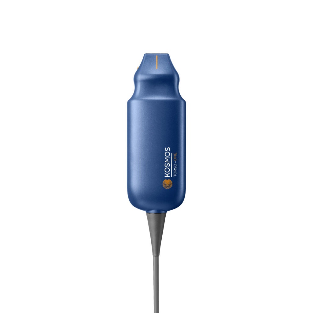
See the heart clearly with the Kosmos Torso-One probe. When making a tough call, you want the cleanest image at the most competitive price.
Conclusion
Hospitalists are increasingly adopting point-of-care ultrasound technology due to its ability to provide immediate bedside diagnostics, significantly improving the efficiency of patient care.
The portability and advanced AI-powered capabilities of Kosmos enable clinicians to make accurate assessments and aid in guiding clinical decisions without the delay of traditional imaging services.
Whether you’re managing heart failure, following a pericardial effusion, or monitoring an aortic aneurysm, Kosmos’ capabilities ensure you capture accurate windows, so you can make better decisions when your patient needs you the most.
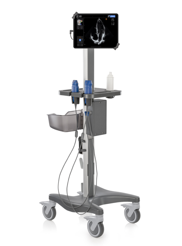
New to ultrasound, or interested in learning more? Our team would be happy to schedule a demo with you. Kosmos Plus allows you to replace your traditional POCUS cart for a fraction of the cost.
Citations
- Chelikam, N., Vyas, A., Desai, R., Khan, N., Raol, K., Kavarthapu, A., Kamani, P., Ibrahim, G., Madireddy, S., Pothuru, S., Shah, P., & Patel, U. K. (2023). Past and Present of Point-of-Care Ultrasound (PoCUS): A Narrative Review. Cureus, 15(12), e50155.
- Dadon, Z., Carasso, S., & Gottlieb, S. (2023). The Role of Hand-Held Cardiac Ultrasound in Patients with COVID-19. Biomedicines, 11(2), 239.
- Singam, N. S. V., Tabi, M., Wiley, B., Anavekar, N., & Jentzer, J. (2023). Echocardiographic findings in cardiogenic shock due to acute myocardial infarction versus heart failure. International journal of cardiology, 384, 38–47.
- Chandraratna, P. A., Mohar, D. S., & Sidarous, P. F. (2014). Role of echocardiography in the treatment of cardiac tamponade. Echocardiography (Mount Kisco, N.Y.), 31(7), 899–910.
- Money, D. B., Mehio, M., Scoma, C., & Gupta, S. (2023). Cardiac Point-of-Care Ultrasound (P.O.C.U.S.) Utilization for Hospitalists in the Assessment of Patients with Cardiac Complaints: An Educational Overview. Journal of community hospital internal medicine perspectives, 13(4), 1–8.
