Peritonsillar Abscess:
The Clinical Utility of Ultrasound
For any clinician, the patient with a suspected peritonsillar abscess (PTA) presents a relatively common but critical diagnostic challenge. The classic presentation of fever, severe odynophagia, trismus, and a “hot potato” voice is often clear, but the key clinical question remains: is this a drainable abscess or simply phlegmon/cellulitis? Historically, this decision point led down one of three paths: a risky blind needle aspiration, a trip to the CT scanner, or a consult for empiric drainage.

Today, point-of-care ultrasound (POCUS) offers a more refined, safer, and efficient fourth path, fundamentally challenging the routine use of CT for uncomplicated cases. For the clinician who may not yet have incorporated this into their workflow, it’s worth examining the evidence.
Table of Contents
The Limitations of Traditional Modalities
While the CT scan has long been considered the “gold standard” for deep neck space infections, its routine use for suspected PTA comes with significant drawbacks.1 The primary demographic for PTA is young adults, a population in whom we strive to limit cumulative ionizing radiation exposure. Furthermore, a CT scan may require IV contrast, has a higher cost burden, and may introduce a delay in care by moving the patient out of the department for imaging, only to answer a question that can often be resolved at the bedside with ultrasound.
Likewise, relying solely on physical exam is fraught with uncertainty, and landmark-based “blind” needle aspirations carry a well-documented risk of failure (if only cellulitis is present) and potential for catastrophic injury to the nearby carotid artery.2
The Evidence for POCUS
Ultrasound directly addresses the shortcomings of the traditional approach by providing rapid, non-invasive, and accurate anatomical information at the bedside.
- Diagnostic Accuracy: Evidence has established the high sensitivity and specificity of ultrasound for PTA, which is comparable to CT for the specific purpose of identifying a drainable fluid collection. Most importantly, POCUS excels at the primary clinical task: differentiating an organized, hypoechoic abscess from the more echogenic, cobblestone appearance of cellulitis. This alone can prevent a significant number of unnecessary drainage procedures.
- Procedural Safety and Success: The paramount advantage of POCUS is its use for real-time procedural guidance. An ultrasound-guided needle aspiration transforms the procedure from a blind attempt into a precise, targeted intervention. Clinicians can visualize the needle tip entering the collection in real-time, confirming its location while simultaneously visualizing and avoiding any critical anatomical areas such as the carotid artery. This not only increases the success rate of aspiration but also adds a profound layer of patient safety.
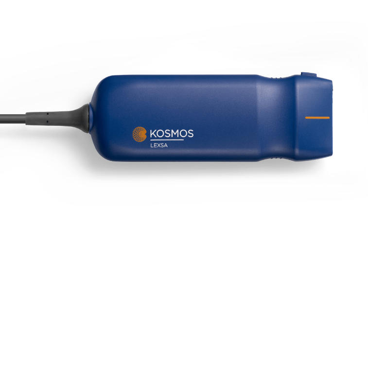
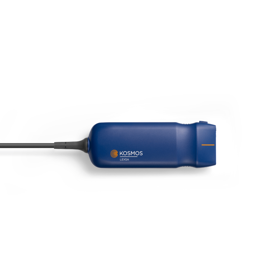
- Workflow Efficiency: Using either a submandibular approach with a linear probe or an intraoral approach with an endocavitary probe, a diagnosis can be confirmed in minutes. This accelerates the decision to drain, treat with antibiotics alone, or consult a specialist, ultimately leading to faster patient disposition.
Re-evaluating the Specialist Consult and the CT Request
The request from an ENT consultant for a CT scan after a positive ultrasound may be rooted in a pre-POCUS era of training, where CT was the only modality to visualize anatomy beneath the mucosal surface, and as such, CT has no role in solely discerning cellulitis from PTA. However, a modern, evidence-based approach calls for risk stratification.
A CT scan remains the indispensable tool for cases with “red flag” features suggesting a more extensive deep neck space infection. These features include significant neck swelling, airway compromise, or suspicion of spread to the parapharyngeal or retropharyngeal spaces. In these complex cases, the global view provided by a CT is non-negotiable for surgical planning.
However, in a patient with a classic, uncomplicated PTA confirmed by ultrasound, a subsequent CT offers little additional information to guide the immediate management (needle aspiration) and can be seen as a medically unnecessary step.
A Modern Algorithm
For the patient with suspected PTA, an evidence-based algorithm should prioritize POCUS:
- Clinical Suspicion: Patient presents with symptoms suggestive of PTA.
- POCUS First: Perform a bedside ultrasound to confirm or exclude a drainable abscess.
- Disposition:
- Cellulitis: Treat with antibiotics.
- Abscess: Perform ultrasound-guided needle aspiration followed with subsequent treatment
- Consult & CT: Reserve CT scans and specialist consultation for patients with inconclusive ultrasounds, failed aspiration, or clinical signs of a complicated deep neck space infection.
By integrating POCUS as the primary imaging modality for suspected uncomplicated PTA, clinicians can make more accurate diagnoses, perform safer procedures, and reduce unnecessary radiation and healthcare costs, representing a clear evolution in the standard of care.
References
- Patel KS, Ahmad S, O’leary G, Michel M. The role of computed tomography in the management of peritonsillar abscess. Otolaryngol Head Neck Surg. 1992;107(6):733-736. doi:10.1177/019459989210700603
- Chang BA, Thamboo A, Burton MJ, Diamond C, Nunez DA. Needle aspiration versus incision and drainage for the treatment of peritonsillar abscess. Cochrane Database Syst Rev. 2016;12(12):CD006287. doi:10.1002/14651858.CD006287.pub4


