What You Need to Know Before Using a Handheld Ultrasound
Point-of-care ultrasound (POCUS) has revolutionized how healthcare providers deliver bedside imaging, offering unparalleled convenience and portability. Handheld ultrasound devices are now widely used across specialties for their compact design and real-time imaging capabilities (1). However, these devices have limitations that may impact clinical workflows and diagnostic efficiency (2). This article explores the potential drawbacks of handheld ultrasound devices and highlights a practical solution in Kosmos.
Key Takeaways:
- Handheld ultrasound devices offer transformative portability and accessibility but are hindered by challenges such as limited battery life, ergonomic inefficiencies, and portability concerns.
- Common issues include the difficulty of balancing paired devices like tablets or phones during use and the risk of misplacing critical components like probes.
- Kosmos provides an innovative solution, overcoming these limitations with enhanced battery life, integrated design, and optimized usability for clinical workflows.
Table of contents
- What You Need to Know Before Using a Handheld Ultrasound
Understanding Handheld Ultrasound and POCUS
What is POCUS?
Point-of-care ultrasound (POCUS) has become a cornerstone of modern bedside diagnostics and procedural guidance. POCUS refers to the use of portable ultrasound devices by healthcare providers to perform focused imaging at the patient’s location, eliminating the need for larger, stationary systems. This approach allows clinicians to assess, diagnose, and guide treatments in real-time, making it invaluable in emergency medicine, critical care, internal medicine, and beyond.
Handheld ultrasound devices are integral to the POCUS revolution. Compact, portable, and user-friendly, these devices empower clinicians to perform immediate imaging at the bedside or in remote settings, enhancing patient outcomes by reducing delays in diagnosis and intervention.
POCUS applications span a broad spectrum of clinical scenarios, including (3-5):
- Focused cardiac ultrasound (FoCUS): Evaluating cardiac function, detecting pericardial effusion, or assessing volume status (5).
- Lung imaging: Identifying pneumothorax, pulmonary edema, or pleural effusions (4).
- Abdominal assessments: Diagnosing free fluid, gallstones, or hydronephrosis (6).
- Vascular access guidance: Ensuring accurate placement of central or peripheral lines (7).
The Rise of Handheld Ultrasound
Portability and Accessibility
Handheld portable ultrasound equipment marks a significant shift in medical imaging. Traditional ultrasound systems are bulky and require designated rooms, limiting their availability. In contrast, handheld devices are small enough to fit in a pocket, enabling seamless integration into clinical workflows (8).
Their accessibility extends beyond hospital walls. Handheld ultrasounds are invaluable in remote clinics, resource-limited settings, or during fieldwork in disaster zones, providing critical imaging where conventional equipment is unavailable (9).
Applications in Clinical Scenarios
Handheld ultrasounds have demonstrated utility across diverse clinical contexts. For example:
- Emergency Medicine: Rapidly assessing trauma patients for internal bleeding (10).
- Critical Care: Monitoring fluid responsiveness and guiding interventions (11).
- Primary Care: Screening for common conditions like abdominal aortic aneurysms or kidney stones (12).
- Sports Medicine: Evaluating musculoskeletal injuries on the field (13).
This versatility cements handheld ultrasound as a tool of choice for clinicians across specialties.
Considerations for Healthcare Providers Using Handheld Ultrasounds
Training and Credentialing
Training Needs
Although handheld ultrasounds are designed for ease of use, proper training is essential. Scanning techniques and image interpretation are skill-based competencies that require hands-on experience. Training programs, workshops, and certifications can help clinicians optimize their use of these devices and ensure diagnostic accuracy (14).
Credentialing Requirements
Credentialing ensures that clinicians using handheld ultrasounds meet standardized safety and quality benchmarks. Institutions should establish protocols to credential users, minimizing risks and improving patient outcomes (15).
Safety Protocols
Overheating Warnings
Handheld ultrasounds, like any electronic device, can generate heat during prolonged use. Manufacturers must incorporate safety mechanisms to prevent overheating and protect both patients and providers (16).
Guidelines for Use
Handheld ultrasounds must adhere to clinical guidelines for their specific applications. Whether used for diagnosis or procedural guidance, adherence to evidence-based practices ensures safe and effective imaging (16).
Image Quality
Comparative Image Clarity
Handheld ultrasounds often produce lower-resolution images compared to larger systems. This limitation may affect diagnostic precision, particularly for detailed assessments such as cardiac function or small organ pathology (17).
Calibration Needs
To achieve optimal imaging, clinicians must adjust parameters such as gain (image brightness) and frequency (depth) based on the body part being examined. Regular calibration is crucial for maintaining image quality (18).
Disinfection Requirements
Rigorous Cleaning Protocols
Given the close contact with patients, handheld ultrasound probes and devices must undergo thorough disinfection between uses. Protocols should align with infection control standards, especially in environments with high patient turnover or contagious diseases (18).
Clinical Documentation and Billing
Charting Results
Accurate documentation of ultrasound findings in the patient’s medical record is essential. This practice supports continuity of care, informs treatment decisions, and provides legal accountability.
Billing and Reimbursement
Handheld ultrasound exams, when clinically indicated, should be eligible for reimbursement. Providers must familiarize themselves with billing codes and criteria to ensure compliance and financial viability.
Hazards and Limitations of Handheld Ultrasounds
Battery Life Concerns
Limited Lifespan
Handheld ultrasound devices are often battery-powered, limiting their use during extended sessions or high patient volumes. Frequent recharging can interrupt workflows and reduce efficiency (19,20).
Power Dependency
Some handheld devices rely on smartphones or tablets for power, draining the paired device’s battery. This dependency can lead to unexpected interruptions during critical moments (19,20).
Ergonomic and Workflow Challenges (21)
Design Limitations
The absence of built-in stands or docking systems forces providers to balance tablets or phones precariously on patients or hold them during scanning. This lack of ergonomics can lead to physical strain and workflow inefficiencies.
Increased Risk of Damage
High-pressure clinical environments increase the likelihood of accidental drops or damage to handheld devices and their paired components.
Risk of Losing Components
Small Components at Risk
Handheld systems typically include small, detachable parts such as probes or cables. Misplacing these components can disrupt workflows and require costly replacements.
Storage Solutions
To address this issue, devices should feature integrated storage or secure docking options to minimize the risk of loss.
Additional Considerations for Handheld Ultrasounds
Focused vs. Comprehensive Exams:
While suitable for focused exams (e.g., cardiac or lung imaging), some handheld devices lack advanced capabilities, such as spectral Doppler, limiting their utility for comprehensive diagnostics.
Avoiding False Findings:
Inappropriate use of handheld ultrasounds can lead to false findings. Providers must exercise clinical judgment and limit usage to situations where these devices are necessary.
Comprehensive Diagnostic Use:
Devices should support both focused imaging and full diagnostic exams to maximize their clinical utility.
Why Kosmos Offers a More Robust Solution
Enhanced Battery Life and Efficiency
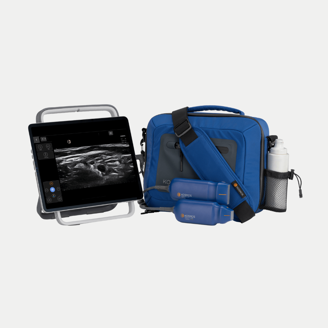
Kosmos addresses the challenge of limited battery life, allowing continuous operation without reliance on external devices for power, offering the ability to scan while plugged into the wall. In addition, with Kosmos Link, users can get up to 8 hours of battery life on a single charge.
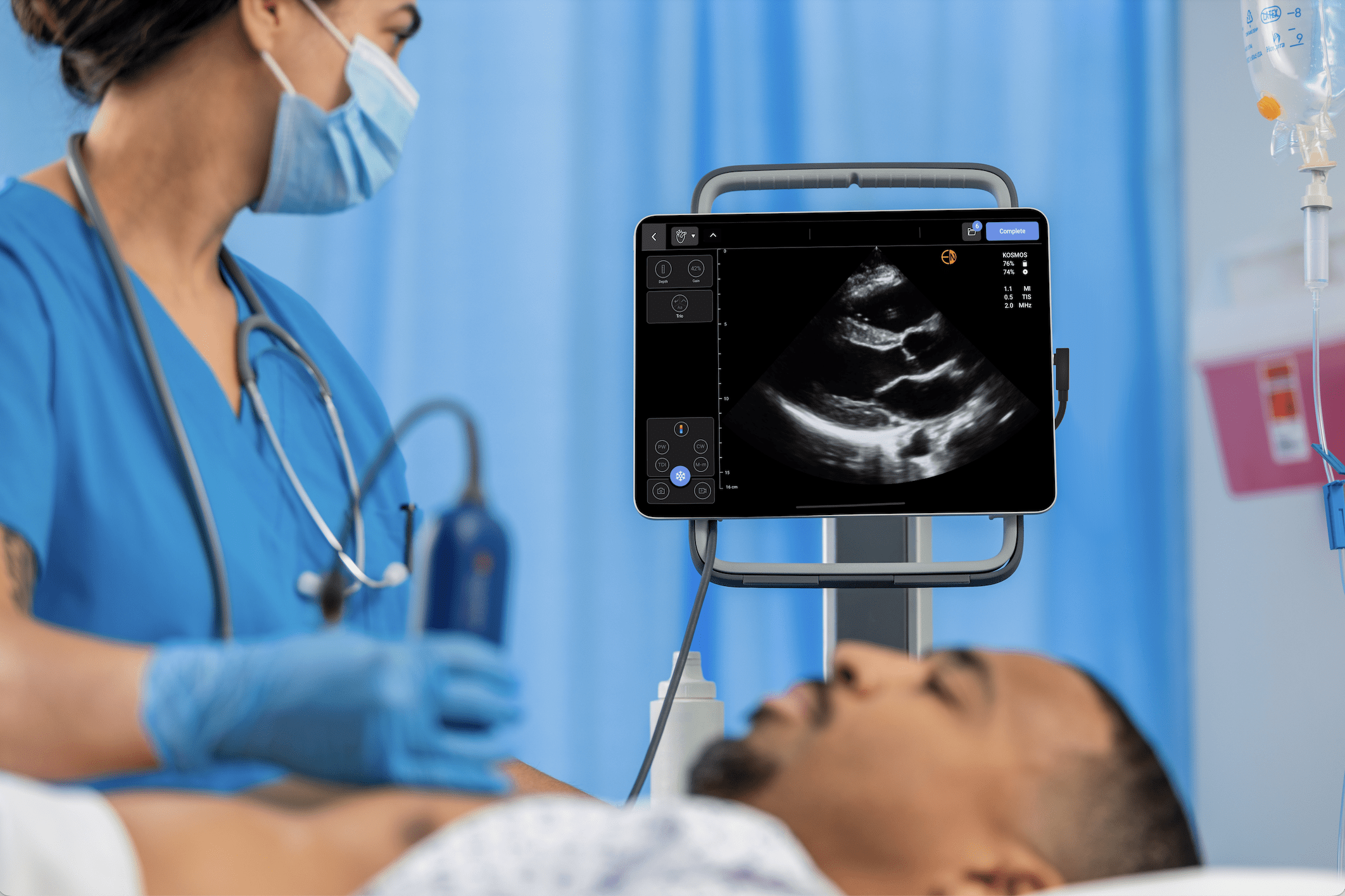
Improved Design and Portability
With an integrated stand and ergonomic design, Kosmos eliminates the need to balance devices precariously, enhancing user experience and reducing risks.
Optimized for Clinical Use
Kosmos is tailored to the demands of clinical environments, offering robust features that ensure safe, efficient imaging without interruptions.
Conclusion
Handheld ultrasound devices have transformed diagnostic and procedural imaging, offering portability and accessibility across diverse clinical settings. However, limitations such as battery life, image quality, and ergonomic challenges must be addressed to optimize their use. Kosmos by EchoNous provides an innovative solution that overcomes these obstacles with superior design and functionality.
References
1. Fraleigh, C. D., et al. (2022). Point-of-care ultrasound: An emerging clinical tool to enhance physical assessment. The Nurse Practitioner, 47(8), 14. PMID: 35877142
2. Elhassan, M. G., et al. (n.d.). Point-of-Care Ultrasonography in Internal Medicine: Limitations and Pitfalls for Novice Users. Cureus, 15(8), e43655.PMID: 37600433
3. Spampinato, M. D., et al. (2024). Diagnostic accuracy of Point Of Care UltraSound (POCUS) in clinical practice: A retrospective, emergency department-based study. Journal of Clinical Ultrasound, 52(3), 255–264. PMID: 38059395
4. Farrell, C., et al. (2024). Exploring the Use of Lung Ultrasonography to Assess Cardiac Surgery Patients: A Scoping Review. Journal of Diagnostic Medical Sonography, 40(1), 79–99.
5. Kalagara, H., et al. (2022). Point-of-Care Ultrasound (POCUS) for the Cardiothoracic Anesthesiologist. Journal of Cardiothoracic and Vascular Anesthesia, 36(4), 1132–1147. PMID: 33563532
6. Khan, M. A. B., et al. (2020). Point-of-care ultrasound for the acute abdomen in primary health care. Turkish Journal of Emergency Medicine, 20(1), 1–11. PMID: 32355895
7. Franco‐Sadud, R., et al. (2019). Recommendations on the Use of Ultrasound Guidance for Central and Peripheral Vascular Access in Adults: A Position Statement of the Society of Hospital Medicine. Journal of Hospital Medicine, 14(9), E1–E22. PMID: 31561287
8. Cardim, N., et al. (2019). The use of handheld ultrasound devices: A position statement of the European Association of Cardiovascular Imaging (2018 update). European Heart Journal. Cardiovascular Imaging, 20(3), 245–252. PMID: 30351358
9. Ienghong, K., et al. (2022). The Utilization of Handheld Ultrasound Devices in a Prehospital Setting. Prehospital and Disaster Medicine, 37(3), 355–359. PMID: 35435155
10. Gao, X., et al. (2023). Progress in the Application of Portable Ultrasound Combined with Artificial Intelligence in Pre-Hospital Emergency and Disaster Sites. Diagnostics, 13(21), 3388. PMID: 37958284
11. Lau, Y. H., et al. (2022). Point-of-care ultrasound for critically-ill patients: A mini-review of key diagnostic features and protocols. World Journal of Critical Care Medicine, 11(2), 70–84. PMID: 35433316
12. Sisó-Almirall, A., et al. (2017). Abdominal aortic aneurysm screening program using hand-held ultrasound in primary healthcare. PloS One, 12(4), e0176877. PMID: 28453577
13. Sofka, C. M. (2004). Ultrasound in sports medicine. Seminars in Musculoskeletal Radiology, 8(1), 17–27. PMID: 30928083
14. Wilkinson, J. N.,et al. (2021). Handheld ultrasound in training – The future is getting smaller! Journal of the Intensive Care Society, 22(3), 220–229. PMID: 34422105
15. Selame, L. A., et al. (2025). Point-of-Care Ultrasound Competency, Credentialing and Policies. Medical Clinics of North America, 109(1), 285–297. PMID: 39567098
16. Hsieh, A., et al. (2022). Handheld Point-of-Care Ultrasound: Safety Considerations for Creating Guidelines. Journal of Intensive Care Medicine, 37(9), 1146–1151. PMID: 35118909
17. Salimi, N., et al. (n.d.). Ultrasound Image Quality Comparison Between a Handheld Ultrasound Transducer and Mid-Range Ultrasound Machine. POCUS Journal, 7(1), 154–159. PMID: 36896280
18. Ferrara, G., et al. (2024). Optimizing Ultrasound Probe Disinfection for Healthcare-Associated Infection Control: A Comparative Analysis of Disinfectant Efficacy. Microorganisms, 12(12), 2394. PMID: 39770597
19. Roy, S., et al. (2019). Chemical Heat Packs as an Intervention to Prolong Ultrasound Battery Runtime. Wilderness & Environmental Medicine, 30(2), 186–190. PMID: 31056373
20. Choi, H. (2019). Prelinearized Class-B Power Amplifier for Piezoelectric Transducers and Portable Ultrasound Systems. Sensors, 19(2), Article 2. PMID: 30642060
21. Baker, J. P., et al. (2013). The Importance of an Ergonomic Workstation to Practicing Sonographers. Journal of Ultrasound in Medicine, 32(8), 1363–1375. PMID: 23887945