Cardiac Ultrasound Machines in 2026: Why the Best Choice Isn’t Necessarily a Traditional Cart
For most of the last two decades, a cardiac ultrasound machine meant more or less one thing: a cart in the echo lab, six figures of capital, scheduled appointments, and a physician trained to ASE Level II or III or RDCS/RCS/ACS.
That machine still matters. It may also be the wrong answer for an increasing share of the cardiac questions clinicians need answered every shift.
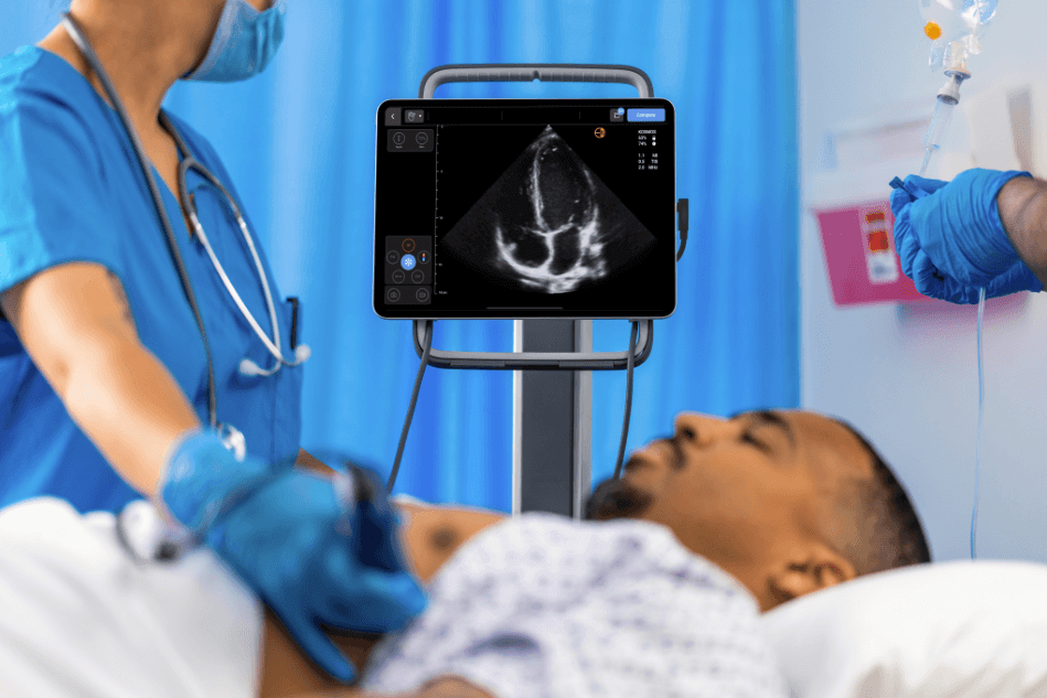
Heart failure rounds don’t exactly happen in the echo lab. Bedside evaluation of suspected aortic stenosis in a busy outpatient clinic does not happen in the echo lab either. Neither does the tamponade rule-out at 3 a.m., the troponin trend on the floor, or the post-TAVI gradient check at the bedside.
For all of those, the right cardiac ultrasound machine in 2026 is the one in your hand when you need it. The question is whether an ultraportable or handheld device can actually deliver cardiac answers, or whether it is just a screening stand-in.
This piece is for cardiologists, hospitalists, intensivists, and the procurement leaders sizing the next cardiac POCUS purchase. We will walk through what a serious cardiac ultrasound machine has to do, where most handhelds fall short, and how the Kosmos platform with the Torso-One phased array probe closes that gap with Continuous Wave Doppler and an AI cardiac workflow that has been studied against standard transthoracic echocardiography and cardiac MRI.1,2
Table of Contents
- Cardiac Ultrasound Machines in 2026: Why the Best Choice Isn’t Necessarily a Traditional Cart
- What a cardiac ultrasound machine actually has to do
- Continuous Wave Doppler, and why most handhelds don’t have it
- AI workflows that compress a measurement into seconds
- What it costs, and what comes with it
- Where Kosmos fits, and where it does not
- Putting it in your hand
- References
- Q&A: Cardiac POCUS Devices
What a cardiac ultrasound machine actually has to do
A cardiac POCUS exam at the bedside is not a stripped-down echo. Cardiac POCUS informs the same clinical decisions in a time-sensitive bedside manner and helps determine when comprehensive consultative echo is needed.3
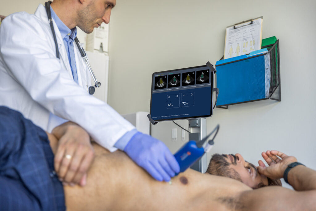
Is the ejection fraction reduced or preserved? How is the global motion? Is there pericardial effusion, or evidence of tamponade? Is the aortic valve gradient consistent with severe stenosis? Is there a regional wall motion abnormality after the troponin bumped? The machine that answers those questions has to do four things well.
- It has to image well. Experts consensus has clarified that the image quality is the most critical characteristic, and that if adequate image quality cannot be obtained, the handheld is “not worth having”.4 That means a phased array probe with a real piezoelectric crystal stack, not a low-cost CMUT transducer that washes out in the apical windows of a larger patient.4 Image quality is where most low-end handhelds quietly give up.
- It has to have the right Doppler capabilities. Color and Pulsed Wave come standard on many devices. Continuous Wave (CW) Doppler does not. CW is the only modality that can help clinicians resolve the high-velocity/mean gradient signals across stenotic or prosthetic valves.5 Without it, a clinician can use a handheld to describe a stenosis but would be unable to grade one, particularly for the Aortic Valve.6
- It has to quantify. Eyeballing LVEF is faster than a Simpson’s biplane, and an experienced cardiologist’s eyeball can be as accurate if not better, with data showing good correlation.7,8,9 Similarly, even non-cardiologists such as ED physicians or intensivists have shown good sensitivity for severe dysfunction.2,17 But payer coverage, guideline-directed medical therapy in heart failure, and longitudinal monitoring all depend on a number, not an impression. A cardiac ultrasound machine that cannot help a clinician produce a quantitative LVEF on demand is incomplete for the bedside use case.
- It has to fit the workflow. Cart-based echocardiography is brilliant in the lab. It is decidedly less useful during rounds. Bedside POCUS can earn its place if the time from “I want a look at the heart” to a usable image is quick and convenient. That is a software and ergonomics problem as much as a hardware one.
Here’s another way of thinking about the buying criteria: image quality good enough that you can trust it, a full Doppler suite including CW, quantitative AI workflows that help clinicians get results in seconds, and a form factor that travels.
The cardiac exams Kosmos is built for
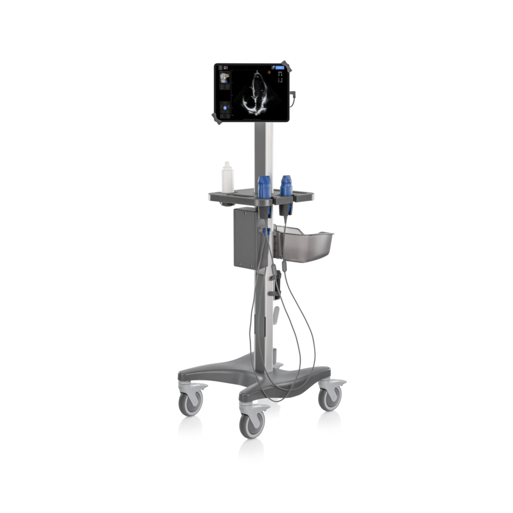
Kosmos, in Mobile and Plus configurations, pairs an iPad-based display with the Torso-One phased array probe, a piezoelectric crystal transducer designed for cardiac, lung, and abdominal imaging on a single probe. In practice, one device covers most of the cardiac questions that arise outside the echo lab.
The platform can help the clinician produce quantitative values, such as LVEF, that maps cleanly onto the HFrEF, HFmrEF, and HFpEF classification used in current guidelines.8
The same exam picks up mitral regurgitation, right ventricular dysfunction, and IVC distention that could change the patient’s management plan.
For valvular heart disease, CW Doppler helps clinicians unlock aortic and mitral gradients at the bedside. A 2025 prospective validation enrolling 101 patients at Hospital Universitario La Paz in Madrid compared handheld Kosmos measurements taken at the bedside to a high-end echo cart, with mean aortic valve gradient as the primary endpoint.
The handheld correctly identified severe aortic stenosis in more than 90 percent of cases.11
For a screening tool deployed in a primary care clinic, a community cardiology office, or the hallway outside the cath lab, that is a clinically meaningful number.
For pericardial monitoring, daily rounds with a handheld may help change the “let us order another echo” reflex. Effusion volume, circumferentiality, and right-sided collapse and IVC plethora that mark tamponade physiology are all visible to a clinician with a Kosmos ultrasound system in a few minutes. A trend over consecutive days may help identify a patient sliding toward tamponade before they get there or those that need escalation to more comprehensive studies.
For post-MI follow-up, regional wall motion abnormalities and serial LVEF measurements are exactly the kind of repeated assessment a cart-based echo schedule cannot accommodate. A handheld ultrasound at the bedside makes the question routine.
For undifferentiated dyspnea, switching between cardiac and lung windows on the same probe lets a single bedside scan can help differentiate decompensated heart failure from pneumonia, pleural effusion, or COPD exacerbation in the same minute. The Auto Preset feature on Kosmos handles the imaging switch automatically when the probe moves from chest to abdomen, so the clinician does not need to stop and change settings.
Continuous Wave Doppler, and why most handhelds don’t have it
The CW Doppler gap may be one of the most consequential capability differences in the handheld market, and it is also the one that is hardest to find in a spec sheet comparison. CW measures velocity along the entire length of the ultrasound beam, which is what allows it to resolve the high-velocity jets that Pulsed Wave Doppler cannot, because of aliasing limits.
Aortic stenosis severity depends on the peak velocity across the valve. So does post-TAVI gradient assessment. So does quantification of mitral or tricuspid regurgitation severity through the peak regurgitant jet velocity. None of those measurements is possible without CW.6
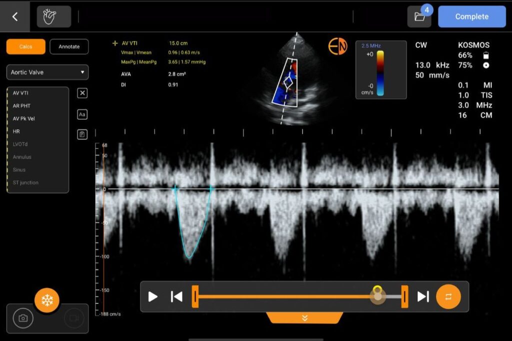
Most handheld ultrasound devices in the current generation do not offer Continuous Wave Doppler.13,14 Kosmos does, through the Torso-One phased array probe. That single capability is what lets a handheld cross from screening into quantitative valvular assessment.
Dr. Jon Zubiaur Zamacola, an interventional cardiology fellow at La Paz who led the AS validation, framed it directly: “Kosmos stood out because it was the first portable device we found that allowed us to perform Continuous Wave Doppler measurements that were previously only available in full-sized echocardiography machines.”11
For aortic stenosis monitoring, TAVI follow-up, and any clinical scenario where valvular hemodynamics matters, the absence of CW on a handheld is a hard ceiling. The presence of CW removes it.
AI workflows that compress a measurement into seconds
Quantitative cardiac measurements have traditionally cost time. Tracing endocardial borders for a biplane Simpson’s LVEF on a cart system takes two to three minutes of dedicated work, which is one of the reasons it is often skipped in favor of a visual estimate.10 AI-assisted measurement changes that math.
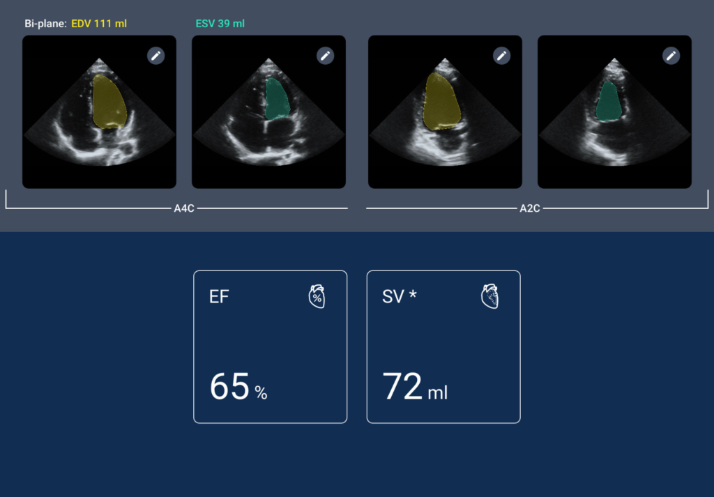
On Kosmos, the Auto EF workflow can run in under twenty seconds.12 The user acquires apical four-chamber and apical two-chamber clips, and and the AI then identifies end-diastole and end-systole, traces the endocardial borders, and proposes a biplane Simpson’s LVEF, which the clinician reviews and confirms.
A 2023 prospective multicenter study published in npj Digital Medicine compared this workflow to formal transthoracic echocardiography and found an intraclass correlation of 0.904 in the overall cohort. The ICC for novice users reached 0.921, slightly higher than the experienced group, which is the kind of result that points at meaningful contributions from AI rather than just labeling existing expertise.2
A 2025 single-center Journal of Clinical Medicine study compared handheld AI-derived LVEF with cardiac MRI in 49 patients, showing near-perfect correlation (r = 0.99), minimal bias of 1.1%, and no significant difference in median LVEF. Larger CMR-referenced studies support the signal: Sveric et al. found AI “Auto-Echo” and Simpson’s method both correlated strongly with CMR in 301 same-day CMR/echo patients (R = 0.89), with AI performing especially well in reduced LVEF, wall-motion abnormalities, and poor image quality, while offering superior reproducibility.15 Mołek-Dziadosz et al. likewise reported strong AI agreement with CMR and expert readers in 118 patients.16 In real-world handheld use, a multicenter AI-POCUS study of 200 patients showed good agreement with standard biplane echo, including ICC 0.81, mean bias around −1.5%, and 85% sensitivity/81% specificity for detecting LVEF <50%.17 Overall, AI-assisted LVEF estimation is increasingly credible as a reproducible adjunct to conventional echocardiography.
Cardiac MRI is the structural gold standard for ventricular function, chiefly volumes and EF. Producing an AI-assisted LVEF at the bedside in under twenty-thirty seconds that shows strong correlation with cardiac MRI is a meaningful shift in what cardiac POCUS can do.
The same cardiac suite includes Auto VTI for stroke volume informing cardiac output and hemodynamic assessments, as well as VExUS based IVC+hepatic/portal/intrarenal venous Doppler workflows alongside associated right-heart hemodynamic assessment, and Auto Doppler, which handles sample gate placement on the inflow tract. Auto Preset switches imaging presets when the probe moves from heart to lung to abdomen, removing the small but constant friction that pulls a clinician out of the scan.
None of this AI requires a subscription from EchoNous. The clinical features that ship with Kosmos remain available without an annual fee, which matters for the total cost of ownership math at the device level and the budget line at the facility level.
What it costs, and what comes with it
A traditional cart-based echocardiography system is a major capital purchase, commonly exceeding six figures, requires dedicated room space, and depends on a sonographer to operate. A Kosmos Plus configuration, with multiple transducers and tablet display in a cart configuration, sits around $20,000, and includes a five-year warranty with no recurring subscription. The economics are not close, with data pointing to meaningfully lower per-exam costs for handheld systems compared with traditional cart-based ones.18,19
For a community cardiology practice, a heart failure clinic, a hospitalist program, or an emergency department adding cardiac POCUS capacity, the relevant comparison is not Kosmos versus a cart. It is Kosmos versus the alternative of referring out, waiting for the next available formal echo slot, and managing the patient in the meantime on physical exam and vital signs alone. The handheld closes that gap.
Where Kosmos fits, and where it does not
It is worth being honest about the boundary. Kosmos is not a replacement for a comprehensive transthoracic echocardiogram performed by a credentialed sonographer in an accredited lab.
Stress echo, transesophageal echocardiography, advanced 3D reconstruction, and the full structural heart workup all stay in the lab. The cart-based system continues to be the right tool for the patient being worked up for surgical aortic valve replacement, the patient with complex congenital anatomy, the patient on the operating table.
What Kosmos fills is the imaging gap between those formal studies. The cardiac assessments that need to happen at the bedside, in the clinic, on rounds, between echo appointments, and in the moments where a decision cannot wait for the schedule. Kosmos answers cardiac questions where they get asked.
Putting it in your hand
If a cardiac ultrasound machine still means a cart in your head, the 2026 version of that question deserves another look. The capability that used to require a six-figure room, a sonographer, and an appointment is increasingly available at the bedside, with Continuous Wave Doppler, AI-assisted LVEF, and the rest of the cardiac workflow on a device that fits in a lab coat pocket.
To see Kosmos Plus or Kosmos Mobile with the Torso-One probe in your own clinical workflow, schedule a demo with our team. We can bring the device, scan the kinds of patients you actually see, and let the imaging make the case.
References
- Bisignani G, Volpe L, Madeo A, Vico R, Bencardino D, De Bonis S. AI-assisted LVEF assessment using a handheld ultrasound device: a single-center comparative study against cardiac magnetic resonance imaging. J Clin Med. 2025;14(13):4708. doi:10.3390/jcm14134708
- Motazedian P, Marbach JA, Prosperi-Porta G, et al. Diagnostic accuracy of point-of-care ultrasound with artificial intelligence-assisted assessment of left ventricular ejection fraction. NPJ Digit Med. 2023;6:201. doi:10.1038/s41746-023-00945-1
- Kirkpatrick JN, Panebianco N, Díaz-Gómez JL, et al. Recommendations for cardiac point-of-care ultrasound nomenclature. J Am Soc Echocardiogr. 2024;37(9):809-819. doi:10.1016/j.echo.2024.05.001
- Perez-Sanchez A, Johnson G, Pucks N, et al. Comparison of 6 handheld ultrasound devices by point-of-care ultrasound experts: a cross-sectional study. Ultrasound J. 2024;16:45. doi:10.1186/s13089-024-00392-3
- Chamsi-Pasha MA, Sengupta PP, Zoghbi WA. Handheld echocardiography: current state and future perspectives. Circulation. 2017;136(22):2178-2188. doi:10.1161/CIRCULATIONAHA.117.026622
- Baumgartner H, Hung J, Bermejo J, et al. Recommendations on the echocardiographic assessment of aortic valve stenosis: a focused update from the European Association of Cardiovascular Imaging and the American Society of Echocardiography. J Am Soc Echocardiogr. 2017;30(4):372-392. doi:10.1016/j.echo.2017.02.009
- Shams P, Goyal A, Makaryus AN. Left ventricular ejection fraction. In: StatPearls. StatPearls Publishing; 2025.
- Heidenreich PA, Bozkurt B, Aguilar D, et al. 2022 AHA/ACC/HFSA guideline for the management of heart failure: a report of the American College of Cardiology/American Heart Association Joint Committee on Clinical Practice Guidelines. Circulation. 2022;145(18):e895-e1032. doi:10.1161/CIR.0000000000001063
- Lang RM, Badano LP, Mor-Avi V, et al. Recommendations for cardiac chamber quantification by echocardiography in adults: an update from the American Society of Echocardiography and the European Association of Cardiovascular Imaging. J Am Soc Echocardiogr. 2015;28(1):1-39. doi:10.1016/j.echo.2014.10.003
- Knackstedt C, Bekkers SCAM, Schummers G, et al. Fully automated versus standard tracking of left ventricular ejection fraction and longitudinal strain: the FAST-EFs multicenter study. J Am Coll Cardiol. 2015;66(13):1456-1466. doi:10.1016/j.jacc.2015.07.052
- Zubiaur J, Salinas P, Nunez-Gil IJ, et al. Validation of a hand-held ultrasound device in the evaluation of aortic stenosis. Int J Cardiovasc Imaging. 2025;41(2):377-385. doi:10.1007/s10554-024-03320-7
- Sachpekidis V, Moustakidis P, Nihoyannopoulos P. Clinical Benchmarking Experience with the Kosmos Platform, a Novel AI-Based Handheld Ultrasound System, After Scanning 1200 Patients in a Tertiary Care Environment, Including 600 Cardiology Consults. EchoNous; 2021.
- Butterfly Network, Inc. Butterfly iQ3 technical specifications. Butterfly Network. Accessed May 2026. https://www.butterflynetwork.com/iq3-specs
- GE HealthCare. Vscan Air CL product details. GE HealthCare. Accessed May 2026. https://www.gehealthcare.com/en-us/products/ultrasound/handheld-ultrasound/vscan-air-sl?
- Sveric KM, Ulbrich S, Dindane Z, Winkler A, Botan R, Mierke J, Trausch A, Heidrich F, Linke A. Improved assessment of left ventricular ejection fraction using artificial intelligence in echocardiography: A comparative analysis with cardiac magnetic resonance imaging. Int J Cardiol. 2024 Jan 1;394:131383. doi: 10.1016/j.ijcard.2023.131383. Epub 2023 Sep 26. PMID: 37757986.
- Mołek-Dziadosz P, Woźniak A, Furman-Niedziejko A, Pieszko K, Szachowicz-Jaworska J, Miszalski-Jamka T, Krupiński M, Dweck MR, Nessler J, Gackowski A. Left ventricular ejection fraction assessment: artificial intelligence compared with echocardiography expert and cardiac magnetic resonance measurements. Pol Arch Intern Med. 2025 Sep 29;135(9):17104. doi: 10.20452/pamw.17104. Epub 2025 Sep 1. PMID: 40888426.
- Kagiyama N, Abe Y, Kusunose K, Kato N, Kaneko T, Murata A, Ota M, Shibayama K, Izumo M, Watanabe H. Multicenter validation study for automated left ventricular ejection fraction assessment using a handheld ultrasound with artificial intelligence. Sci Rep. 2024 Jul 4;14(1):15359. doi: 10.1038/s41598-024-65557-5. PMID: 38965290; PMCID: PMC11224326.
- Stella-Lida Papadopoulou, Foteini Malakoudi, Christina Tsantekidou, Anastasios Papanastasiou, Dimitrios Dionysopoulos, Nikolas Moustakidis, Theofilos Moustakidis, Panagiotis Stafylas, Ioannis Styliadis, Petros Nihoyannopoulos, Areti Triantafyllou, Vasileios Sachpekidis, Diagnostic accuracy and cost-effectiveness of a handheld ultrasound device for cardiac evaluation in noncardiology settings, European Heart Journal – Imaging Methods and Practice, Volume 4, Issue 1, January 2026, qyag047, https://doi.org/10.1093/ehjimp/qyag047
- Trambaiolo P, Papetti F, Posteraro A, Amici E, Piccoli M, Cerquetani E, Pastena G, Gambelli G, Salustri A. A hand-carried cardiac ultrasound device in the outpatient cardiology clinic reduces the need for standard echocardiography. Heart. 2007 Apr;93(4):470-5. doi: 10.1136/hrt.2006.094201. Epub 2006 Aug 29. PMID: 16940393; PMCID: PMC1861486.
Q&A: Cardiac POCUS Devices
Continuous Wave (CW) Doppler is the only modality capable of helping clinicians resolve high-velocity signals across stenotic or prosthetic valves. Without this capability, a clinician can use a handheld device to describe a stenosis but would be unable to grade it, particularly for the Aortic Valve. Most current-generation handheld ultrasound devices lack Continuous Wave Doppler , making its inclusion in devices like Kosmos a significant differentiator for quantitative valvular assessment.
Quantitative cardiac measurements have traditionally cost time, but AI-assisted workflows change that math by helping clinicians acquire results in seconds. On the Kosmos platform, the Auto EF workflow can run in under twenty seconds. The AI identifies end-diastole and end-systole, traces the endocardial borders, and proposes a biplane Simpson’s LVEF, which the clinician then reviews and confirms. Producing an AI-assisted LVEF at the bedside shows a strong correlation with cardiac MRI , which is considered the structural gold standard for ventricular function.
Handheld systems like Kosmos are not a replacement for a comprehensive transthoracic echocardiogram performed by a credentialed sonographer in an accredited lab. Cart-based systems continue to be the right tool for stress echoes, transesophageal echocardiography, advanced 3D reconstruction, and complete structural heart workups. Instead, handheld devices are designed to fill the imaging gap between those formal studies. They provide rapid cardiac assessments that need to happen at the bedside, in the clinic, on rounds, and between scheduled echo appointments.
A traditional cart-based echocardiography system is a major capital purchase that commonly exceeds six figures. In contrast, a Kosmos Plus configuration sits at around $20,000 and includes a five-year warranty with no recurring subscription. Furthermore, Kosmos provides its clinical AI features without an annual subscription fee, lowering the total cost of ownership at the facility level.