Patient: An 82 year old female patient presented to the ER with symptoms and signs of congestive heart failure.

Initial Examination: She had a history of well controlled hypertension treated with beta blockers and diuretics by her primary care physician. On cardiac auscultation a mid-systolic ejection murmur was noted with diminished S2 heart sound. Her jugular veins were dilated and mild lower limb edema was present.

Kosmos POCUS Examination: Bedside examination with KOSMOS revealed severely impaired left ventricular systolic function. The aortic valve was heavily calcified and had severe stenosis. Moderate functional mitral regurgitation was also present. Patient was treated with intravenous diuresis resulting in improvement of her symptoms. A subsequent coronary angiogram revealed no coronary stenoses. She was scheduled for a TAVI procedure.
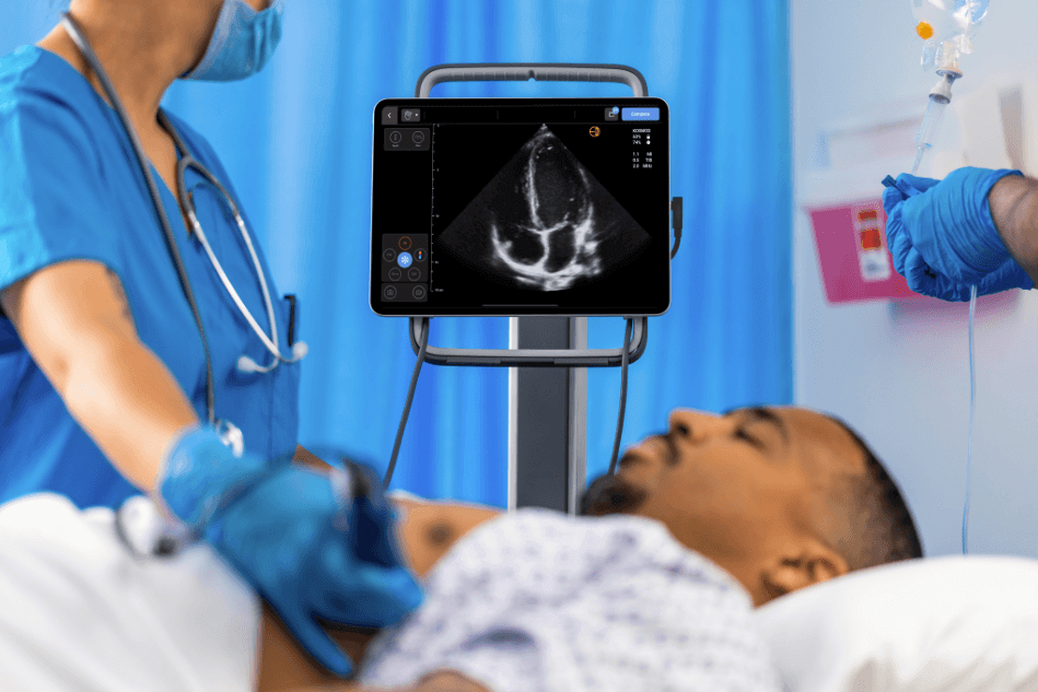
Video 1
PLAX view showing dilated left ventricle with severely impaired overall systolic function. Diffuse hypokinesis of LV walls is seen. The aortic valve is heavily calcified and not opening well. There is symmetric tethering of both mitral valve leaflets due to LV remodeling. There is also mild pericardial effusion.

Video 2
PLAX view with color showing moderate functional mitral regurgitation.

Video 3
PLAX view with color on the aortic valve showing aortic stenosis and mild aortic regurgitation.

Video 4
PSAX view at the level of the papillary muscles showing severely impaired LV systolic function.

Video 5
PSAX view at the level of the aortic valve showing a heavily calcified tricuspid with stenotic behavior.

Video 6
PSAX view at the level of the aortic valve with color showing mild aortic valve regurgitation.

Video 7
A4C view showing dilated LV with severely impaired systolic function. The RV has normal size and systolic function. There is also bi-atrial enlargement. A small pericardial effusion is evident.

Video 8
A2C view showing dilated LV with severely impaired systolic function.

Video 9
A3C view showing dilated LV with severely impaired systolic function. The aortic valve is severely calcified.

Figure 1
Automated EF calculation with KOSMOS AI confirming the visual impression of severely impaired LV systolic function.

Video 10
A4C view with color showing moderate functional mitral regurgitation.

Video 11
CW MR signal from apical four chamber view.

Video 12
A5C view with color showing turbulent flow through the stenosed AV.

Video 13
CW AS signal from A5C view.
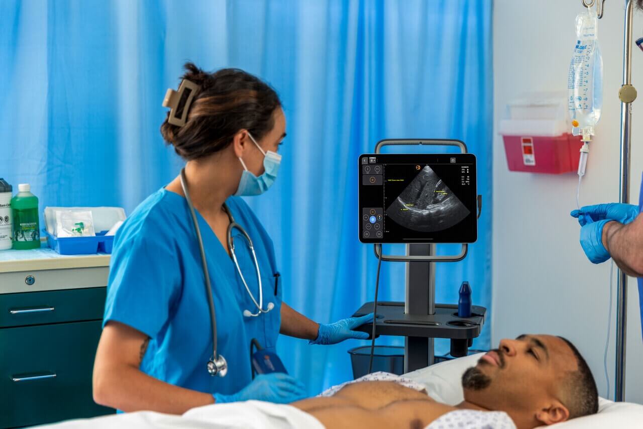
Figure 2
Maximum aortic valve velocity measured at 3,7-3,8 m/s with CW.

Video 14
CW AR signal from A5C view.

Video 15
PW LVOT signal showing reduced forward LV stroke volume
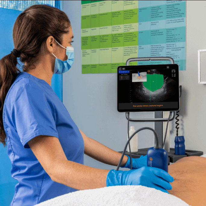
Figure 3
Maximum LVOT velocity measured at 0.54 m/s with PW, suggestive of low flow state.

Video 16
A4C view with color showing mild functional tricuspid regurgitation. There is also color flow inside the RA coming from the IAS, suggestive of a patent PFO with left to right shunt due to increased LA pressure.

Video 17
Subcostal view showing dilated IVC.

  • The Evolution of Ultrasound in Emergency Medicine Practice

    The Evolution of Ultrasound in Emergency Medicine Practice

    The Evolution of Ultrasound in Emergency Medicine Practice Over the past decade, ultrasound has revolutionized emergency medicine, providing real-time bedside diagnostics to guide clinical decisions in time-sensitive scenarios. With this direct access, ultrasound provides the…

    Read More

  • One System for Your Nursing Ultrasound Needs

    One System for Your Nursing Ultrasound Needs

    The Benefits of a Dual-Purpose Ultrasound System for Vascular Access and Bladder Scanning Efficiency and accuracy are central to modern nursing practice, and fundamental bedside procedures such as vascular access and bladder assessment can have…

    Read More

  • What’s Under the Hood of Your Ultrasound Device?

    What’s Under the Hood of Your Ultrasound Device?

    What’s Under the Hood of Your Ultrasound Device? Point-of-care ultrasound (POCUS) has revolutionized bedside diagnostics, allowing clinicians to visualize structures and perform interventions with precision. Portable ultrasound equipment combines advanced technologies, including harmonic imaging and…

    Read More

  • Lung Ultrasound in Acute and Chronic Heart Failure

    Lung Ultrasound in Acute and Chronic Heart Failure

    Lung Ultrasound in Heart Failure Management: Enhancing Diagnosis, Monitoring, and Patient Outcomes in Critical Care Heart failure (HF) represents a significant clinical problem affecting an estimated 60 million people worldwide who require care. It is…

    Read More