A 73 year old male patient presented with right sided heart failure. His ECG revealed atrial fibrillation.

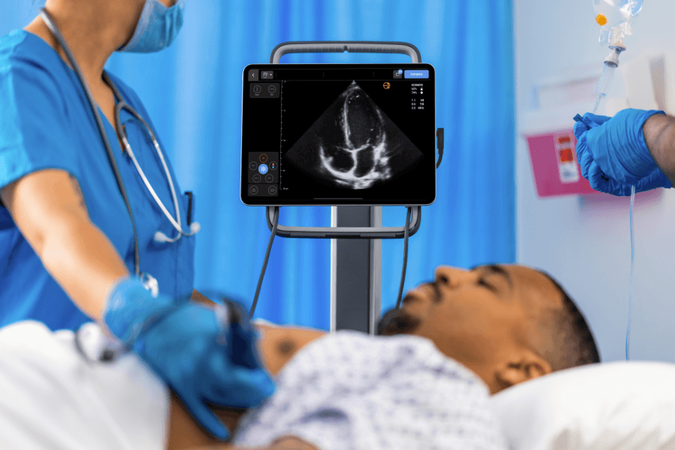
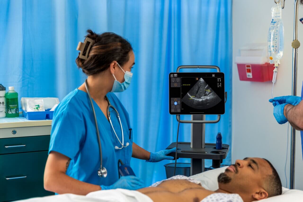
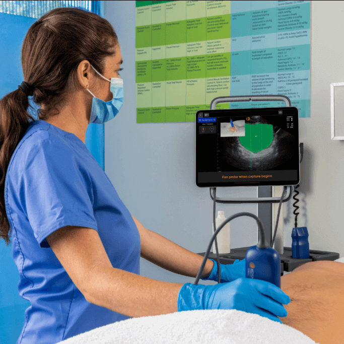
On clinical examination increased jugular venous pressure and a holosystolic murmur was noted. Bedside examination with KOSMOS revealed thickened LV and RV walls with impaired systolic function of both ventricles. All valves were mildly thickened. Mild to moderate mitral regurgitation and significant tricuspid regurgitation were seen. The IVC was dilated and mild pericardial effusion was noted. All these features strongly pointed towards cardiac amyloidosis which was later confirmed with cardiac MRI.

A4C view showing thickened ventricles with impaired systolic function

A4C view showing significant tricuspid regurgitation

CW signal of tricuspid regurgitation

A2C and A3C views showing impaired LV systolic function 01

A2C and A3C views showing impaired LV systolic function 02

A3C view showing mild to moderate mitral regurgitation

Subcostal long axis view showing thickened ventricles with impaired systolic function

Dilated IVC

  • Bowel Ultrasound for Family Physicians

    Bowel Ultrasound for Family Physicians

    Bowel Ultrasound Complete Guide for Family Physicians to Master IBD Care GIT complaints are very common in family medicine. Bloating, diarrhea, and abdominal pain are the main symptoms indicating the presence of an inflammatory bowel…

    Read More

  • What You Need to Know Before Using a Handheld Ultrasound

    What You Need to Know Before Using a Handheld Ultrasound

    What You Need to Know Before Using a Handheld Ultrasound Point-of-care ultrasound (POCUS) has revolutionized how healthcare providers deliver bedside imaging, offering unparalleled convenience and portability. Handheld ultrasound devices are now widely used across specialties…

    Read More

  • Essential ultrasound protocols for the critically ill patient

    Essential ultrasound protocols for the critically ill patient

    Essential Ultrasound Protocols for Critically Ill Patients: EFAST, RUSH, and SHoC-ED In critical care, time isn’t just valuable—it’s everything. When a patient experiences cardiac arrest or shock, there’s no time to lose. Clinicians need instant…

    Read More

  • Ultrasound for Monitoring Diaphragm Dysfunction after Cardiac Surgery

    Ultrasound for Monitoring Diaphragm Dysfunction after Cardiac Surgery

    Ultrasound for Monitoring Diaphragm Dysfunction after Cardiac Surgery Point-of-care ultrasound (POCUS) is gaining a reputation as a bedside diagnostic and monitoring tool in critical patient care. This reputation is mostly connected to its effectiveness, versatility,…

    Read More