Ultrasound Device Options for Vascular Access
Selecting the right ultrasound device and probe is a key factor in the success of ultrasound-guided peripheral IV (PIV) insertion. The market offers a wide range of equipment options, from high-end cart-based systems to compact, handheld units. Choosing the best solution depends on your clinical setting, workflow needs, and patient population. In this article, we’ll explore the essential components of ultrasound equipment used for IV access, with a focus on probe selection, device types, and feature considerations.
Table of Contents
The Role of Equipment in Ultrasound-Guided IV Placement
While operator skill is the most important determinant of success, high-quality imaging tools greatly enhance the ability to visualize superficial veins, track needle advancement in real time, and confirm proper placement. Poor image quality or cumbersome equipment can frustrate workflows and reduce first-pass success. Matching your device to the intended use case—whether in an emergency department, ICU, general ward, or outpatient infusion center—is critical to achieving reliable outcomes.
The success of ultrasound-guided venous access greatly depends on having the appropriate ultrasonography equipment. Basic models function, but better units produce better outcomes.
Selecting the Right Transducer
Imaging Modes for IV Placement
Standard two-dimensional (B-mode) grayscale imaging is generally all that is needed for venous cannulation. Doppler ultrasound is sometimes helpful for vein selection (eg, identifying thrombosis, differentiating vein from an artery.)
B-Mode (2D Imaging)
B-mode is the standard for ultrasound-guided vascular access. It provides a grayscale image of the vessel and surrounding tissue, allowing:
- Real-time visualization of vein lumen and compressibility
- Differentiation of veins from arteries (with compression or pulsatility)
- Needle tracking under dynamic guidance
Doppler Mode
While not required for routine cannulation, Doppler adds valuable flow information.
- Color Doppler overlays flow direction and relative velocity on the grayscale image, helping confirm vessel patency and distinguish arteries from veins when anatomy is unclear.
- Spectral Doppler produces a velocity–time waveform for quantitative flow analysis, more often used in vascular labs than in routine IV placement.
Duplex Imaging
Duplex ultrasound combines B-mode with Doppler (color or spectral), enabling simultaneous visualization of vessel anatomy and flow. However, Doppler sensitivity decreases when the vessel lies at a 90° angle to the transducer. Tilting the probe to optimize the Doppler angle improves signal detection. For most peripheral IV insertions, standard 2D B-mode provides the best temporal resolution and remains the primary imaging method.
Linear Array Probes are the Gold Standard
Ultrasound machines vary in sophistication. While basic models work for ultrasound-guided venous access, higher-quality units produce better results. For vascular access, use a high-frequency (5-15 MHz) linear transducer. It provides high-resolution imaging close to the skin surface. These probes offer excellent resolution for imaging superficial structures like forearm or antecubital fossa veins.
For vein imaging, a linear shape works best because it allows for even compression, which helps differentiate veins from arteries.
- Better needle tip visualization
- Wide field of viewing for vein tracking
- For vein imaging, a linear shape works best because it allows for even compression, which helps differentiate veins from arteries.
Ultrasound System Types
1. Cart-Based Systems
These traditional ultrasound machines provide premium image quality and advanced features but are less portable.
Pros
- Superior resolution and processing power
- Often include Doppler, needle enhancement, and large touchscreens
- Better suited for ICUs, operating rooms, and radiology departments
Cons
- Expensive
- Less mobile, more difficult to maneuver in tight spaces
2. Portable Units
Compact systems that offer a middle ground between functionality and portability.
- Useful in emergency departments, procedural suites, and mobile response teams
- Can include most features of cart-based systems
- Easier to integrate into bedside workflows
3. Handheld Ultrasound Devices
Handheld units connect to tablets or smartphones and deliver performance in a small form factor.
Pros
- Highly portable and lightweight
- Quick boot-up and easy sterilization
- Convenient for bedside IV placement, emergency settings
Cons
- Smaller screens can be harder for needle visualization
- Fewer advanced features than larger units
Tailoring Equipment to Clinical Settings
| Settings | Recommended Device | Why? |
| Emergency Department | Handheld or portable | Fast deployment, mobility, cost-effective |
| ICU | Cart-based or portable | Higher-end features, best image resolution |
| Med/Surg Floors | Handheld or portable | Streamlined workflows, cost-conscious |
| Labor & Delivery | Handheld or portable | Quick vein access in urgent maternal/fetal settings; easy to sterilize |
| Outpatient Infusion | Handheld or portable | Frequent IV placements, rapid turnaround, lower cost |
Key Features to Consider for Vascular Access Ultrasound
When evaluating ultrasound devices for vascular access, clinicians should prioritize features that enhance procedural efficiency, accuracy, and patient safety. Here are the key features to consider, tailored to the capabilities of the Kosmos ultrasound platform:
Exceptional Image Quality for Clear Visualization
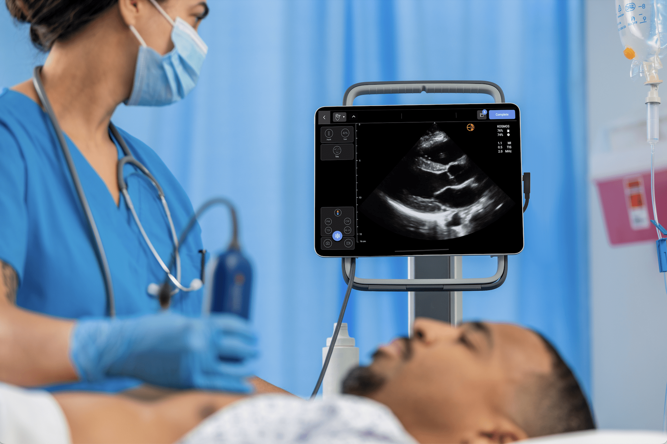
For confident and precise needle placement, superior image quality is paramount. A system with a direct, wired probe connection ensures a stable, interference-free signal, delivering the highest possible image clarity. Look for a device with high-frequency linear probes and advanced compound imaging technology to clearly delineate vessels and surrounding anatomy. The Kosmos Lexsa probe, for instance, provides outstanding, high-resolution images, allowing for precise visualization of needles, veins, arteries, and other structures up to a 10cm depth.
Reliable Portability and Workflow Integration
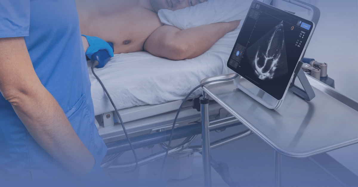
In the fast-paced environment of vascular access, a reliable and portable system is essential. While portable, the Kosmos system utilizes a cabled connection between the probe and the tablet. This design choice intentionally maximizes reliability and durability, eliminating concerns about signal loss or battery issues in the probe itself during critical procedures. The system’s tablet-based platform still offers excellent portability for easy transport between patient rooms and at the point of care.
Intuitive User Interface and Ease of Use
A user-friendly interface is critical for rapid and efficient use during procedures. Look for a device with a responsive touchscreen that allows for quick adjustments to depth, gain, and other imaging parameters with minimal effort. The ability to quickly switch between different presets with a simple tap can save valuable time. The Kosmos platform is known for its user-centric design, featuring a streamlined workflow and an intuitive interface.
Specialized Probes and Advanced Doppler Capabilities
The right probe can make a significant difference in vascular access procedures. A high-frequency linear probe, like the Kosmos Lexsa, is ideal for visualizing superficial vessels with exceptional detail. Additionally, the availability of advanced Doppler features, such as Color Doppler, Color Power Doppler, and Pulsed Wave Doppler, is crucial for easy identification and assessment of vascular structures, further enhancing procedural safety and success.
Durability, Battery Life, and Infection Control
Given the demands of a busy clinical setting, the device’s durability and battery life are important considerations. Look for a device that is drop-tested and has a long-lasting battery in the main tablet unit to ensure it can withstand the rigors of daily use. Furthermore, ensure the device and probes are compatible with standard sterile sheaths and can be easily cleaned with high-level disinfection systems to maintain a safe clinical environment. Kosmos probes are compatible with the Trophon disinfection system, a high-level solution that eliminates pathogens while protecting probe integrity.
Summary
The success of ultrasound-guided IV insertion depends not only on clinician skill but also on choosing the right ultrasound system and probe. High-frequency linear probes remain essential for superficial vein visualization, while handheld and portable devices provide the mobility and speed increasingly demanded across care settings. When selecting a device, consider:
- Clinical environment (emergency, ICU, med/surg, outpatient)
- Frequency of use and patient mix
- Required features (needle enhancement, wireless access)
- Budget and training infrastructure
Well-chosen equipment improves first-pass success, reduces complications, and supports the broader adoption of ultrasound guidance in vascular access protocols.
References
- AIUM Practice Parameter for the Use of Ultrasound to Guide Vascular Access Procedures. J Ultrasound Med. 2019;38(3):E4-E18. doi:10.1002/jum.14954
- Kerforne T, Petitpas F, Frasca D, Goudet V, Robert R, Mimoz O. Ultrasound-guided peripheral venous access in severely ill patients with suspected difficult vascular puncture. Chest. 2012;141(1):279-280. doi:10.1378/chest.11-2054
- Lamperti M, Bodenham AR, Pittiruti M, et al. International evidence-based recommendations on ultrasound-guided vascular access. Intensive Care Med. 2012;38(7):1105-1117. doi:10.1007/s00134-012-2597-x
- Levy JA, Noble VE. Bedside ultrasound in pediatric emergency medicine. Pediatrics 2008; 121:e1404.
- Liu L, Tan Y, Li S, Tian J. “Modified Dynamic Needle Tip Positioning” Short-Axis, Out-of-Plane, Ultrasound-Guided Radial Artery
- Liu YT, Alsaawi A, Bjornsson HM. Ultrasound-guided peripheral venous access: a systematic review of randomized-controlled trials. Eur J Emerg Med. 2014;21(1):18-23. doi:10.1097/MEJ.0b013e328363bebc
- Moore CL, Copel JA. Point-of-care ultrasonography. N Engl J Med. 2011;364(8):749-757. doi:10.1056/NEJMra0909487
- Shokoohi H, Boniface K, McCarthy M, et al. Ultrasound-guided peripheral intravenous access program is associated with a marked reduction in central venous catheter use in noncritically ill emergency department patients. Ann Emerg Med. 2013;61(2):198-203. doi:10.1016/j.annemergmed.2012.09.016
- Stolz LA, Stolz U, Howe C, Farrell IJ, Adhikari S. Ultrasound-guided peripheral venous access: a meta-analysis and systematic review. J Vasc Access. 2015;16(4):321-326. doi:10.5301/jva.5000346
- Ultrasound Guidelines: Emergency, Point-of-Care and Clinical Ultrasound Guidelines in Medicine. Ann Emerg Med. 2017;69(5):e27-e54. doi:10.1016/j.annemergmed.2016.08.457
- Whitson MR, Mayo PH. Ultrasonography in the emergency department. Crit Care. 2016;20(1):227. Published 2016 Aug 15. doi:10.1186/s13054-016-1399-x
- Butterfly Network – Butterfly iQ+ Product Specifications.
- Philips Healthcare. Lumify Portable Ultrasound Product Details.
- GE Healthcare. Vscan Air Overview.
- Kosmos by EchoNous. Product Specifications and Vascular Access Applications.