Retropharyngeal Abscess:
The Evolving Role of Bedside Ultrasound
The diagnosis of a retropharyngeal abscess (RPA) represents a serious challenge in emergency medicine and pediatrics. Primarily affecting children five and younger, retropharyngeal abscess is uncommon but increased in incidence in the 2000’s.1
RPA is a life-threatening deep neck infection that can rapidly lead to airway compromise or spread to the chest (mediastinum). The classic symptoms of fever, neck stiffness, and dysphagia are often inconsistent in this population, where non-specific signs can create a broad differential.
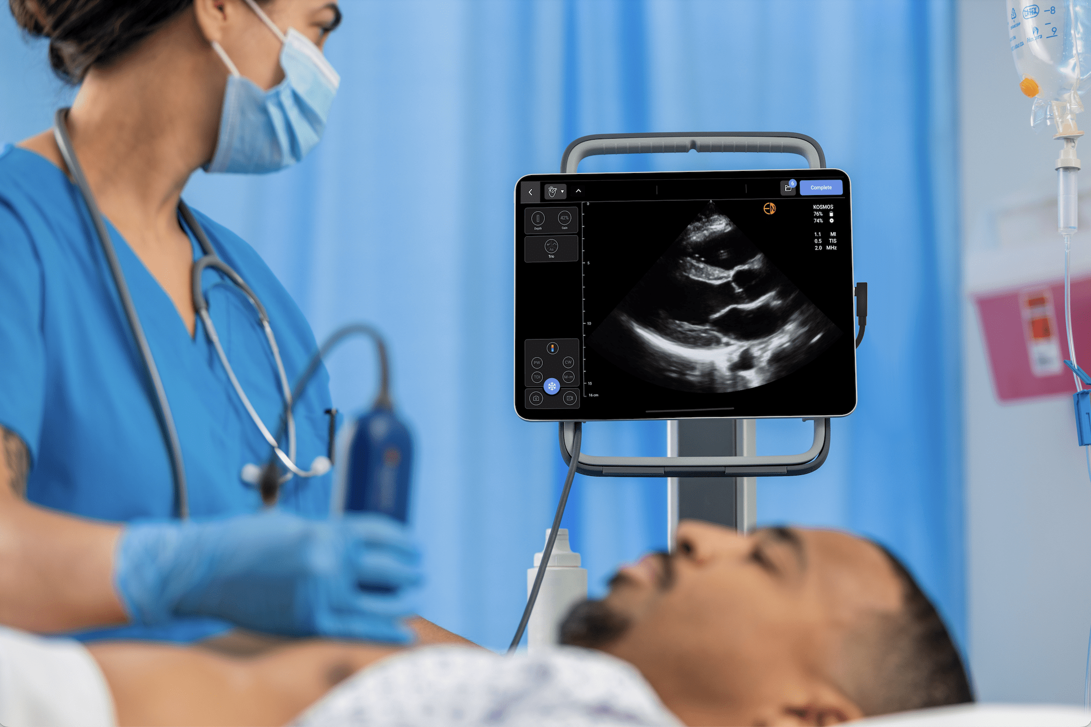
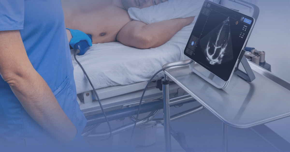
Because the abscess is often not detectable by physical exam alone, advanced imaging becomes essential, and the choice of modality has significant clinical implications. While CT with contrast has been the diagnostic standard, point-of-care ultrasound (POCUS) is emerging as a powerful, radiation-sparing tool to rapidly screen for and diagnose this dangerous condition at the bedside.2
Table of Contents
The Limitations of Traditional Modalities
Historically, the initial imaging test for a suspected RPA was a lateral neck soft tissue radiograph.3 Its utility, however, is notoriously limited. The diagnostic criterion—widening of the prevertebral soft tissues—is highly dependent on patient cooperation, requiring a perfectly positioned neck in full extension during inspiration. In a crying, struggling child, this is nearly impossible to achieve, leading to a high rate of false-positive results and low overall sensitivity.4
Consequently, the contrast-enhanced CT scan became the definitive gold standard. It provides exquisite anatomical detail, confirms the presence of a drainable collection, and delineates any extension into surrounding structures.5
However, this diagnostic certainty comes at a significant cost, particularly in children: a high dose of ionizing radiation and the frequent need for sedation. Sedating a child with a potentially compromised airway carries inherent risks, while the entire process delays a time-sensitive diagnosis. Further, although considered the gold standard, literature has varied on specificity and sensitivity, with evidence showing values as low as 40-50%.6
The Evidence for POCUS
Point-of-care ultrasound offers a compelling alternative as a first-line imaging tool, directly addressing the safety and efficiency concerns of traditional methods.
- Diagnostic Accuracy: Two techniques have been described in the literature.2,4 Mainly, using a high-frequency linear probe in a transverse and longitudinal orientation over the anterior neck, clinicians can directly visualize and measure the prevertebral soft tissue. Studies have demonstrated that ultrasound can match CT for detecting RPA, with some evidence suggesting it is superior to lateral radiography and may approach or exceed the accuracy of CT for identifying a fluid collection.5 It can reliably differentiate the normal, thin prevertebral space from the thickened, edematous appearance of phlegmon or the complex, hypoechoic fluid collection of a mature abscess.
- Safety and Workflow Efficiency: The most profound benefit of POCUS is the complete avoidance of ionizing radiation. It can be performed in minutes at the bedside without removing the patient from a monitored setting and, crucially, avoid the need for sedation. This can allow for the rapid identification of patients who require urgent consultation and a pre-operative CT, while also providing the confidence to rule out the diagnosis in lower-risk patients, potentially preventing a CT scan and hospital admission altogether.
Re-evaluating the CT Scan in the POCUS Era
The role of POCUS is not to completely replace the CT scan, but rather to fundamentally change its position in the diagnostic algorithm.
The CT scan remains the undisputed gold standard for pre-operative surgical planning as well as to confirm or rule out the diagnosis fully.
An ENT or surgical specialist requires the detailed anatomical map a CT provides to assess the full extent of the abscess and its relationship to vital structures before proceeding to the operating room.
The modern role for POCUS, therefore, is as a powerful diagnostic screening test.
- A positive POCUS exam that identifies a fluid collection confirms the diagnosis of RPA. This result justifies the radiation and sedation risks of a subsequent CT scan, which now serves a pre-operative, confirmatory purpose rather than a diagnostic one.
- A negative POCUS exam that shows normal prevertebral soft tissue in a patient with low-to-moderate clinical suspicion has a high negative predictive value. This can enable clinicians to exclude RPA if clinically deemed low risk and avoid the downstream cascade of a CT scan, admission, and consultation.
A POCUS-prioritizing Algorithm for RPA
For the patient with suspected RPA, an evidence-based algorithm should prioritize POCUS:
- Clinical Suspicion: Patient (often pediatric) presents with fever, torticollis, neck pain/stiffness, drooling, or stridor
- POCUS First: Perform a bedside ultrasound of the anterior neck to visualize and measure the prevertebral space
- Disposition:
- Negative POCUS: In a non-toxic appearing patient, this strongly argues against RPA. Consider alternative diagnoses and observation.
- Positive POCUS (Fluid Collection): Confirms RPA. Obtain an urgent ENT consult and proceed with a contrast-enhanced CT scan for pre-operative planning.
- Equivocal POCUS (Phlegmon/Cellulitis): The decision for CT and consultation should be based on the patient’s clinical trajectory and overall degree of suspicion.
By integrating POCUS as the initial imaging modality for suspected RPA, clinicians can significantly reduce radiation exposure in children, avoid the risks of sedation, and streamline the diagnostic pathway for this true airway emergency.
Q&A: Retropharyngeal Abscess and the Role of Bedside Ultrasound
A retropharyngeal abscess is a deep neck space infection located behind the pharynx. It primarily affects children under five and can progress rapidly, leading to airway obstruction or mediastinal spread if not diagnosed promptly.
Children often present with vague or inconsistent symptoms such as fever, neck pain, or difficulty swallowing. Physical examination alone cannot reliably identify an abscess due to the deep location of the infection, making imaging essential.
Lateral neck X-rays have poor sensitivity and are highly position-dependent, often yielding false positives. CT scans offer detailed anatomy but require ionizing radiation and sedation—both risky for children—and can delay urgent management.
POCUS allows clinicians to visualize the prevertebral soft tissue at the bedside. It can identify thickened tissue, phlegmon, or abscess formation within minutes—without radiation or sedation. Studies show its diagnostic accuracy may rival or exceed traditional imaging for detecting fluid collections.
POCUS serves as a first-line screening tool. A positive POCUS confirms the need for CT for surgical planning, while a negative POCUS in a low-risk patient can safely rule out RPA and avoid unnecessary imaging.
– Eliminates radiation exposure
– Avoids sedation in children
– Provides rapid, bedside answers
– Reduces unnecessary CT scans and admissions
– Enhances workflow efficiency and patient safety
Integrating POCUS can reduce unnecessary radiation exposure, expedite diagnosis, and improve patient outcomes by allowing earlier recognition and management of life-threatening infections like RPA.
References
- Woods CR, Cash ED, Smith AM, et al. Retropharyngeal and Parapharyngeal Abscesses Among Children and Adolescents in the United States: Epidemiology and Management Trends, 2003–2012. J Pediatric Infect Dis Soc. 2016;5(3):259-268. doi:10.1093/jpids/piv010
- Scheier E, Shapira-Levy E, Oren S. Point of care ultrasound (POCUS) diagnosis of deep neck space abscess: a case series. J Emerg Med. 2025;71:60-66. doi:10.1016/j.jemermed.2024.10.004
- Brechtelsbauer PB, Garetz SL, Gebarski SS, Bradford CR. Retropharyngeal abscess: pitfalls of plain films and computed tomography. Am J Otolaryngol. 1997;18(4):258-262. doi:10.1016/s0196-0709(97)90006-5
- Malia L, Sivitz A, Chicaiza H. A novel approach: point-of-care ultrasound for the diagnosis of retropharyngeal abscess. Am J Emerg Med. 2021;46:271-275.
- Glasier CM, Stark JE, Jacobs RF, et al. CT and ultrasound imaging of retropharyngeal abscesses in children. AJNR Am J Neuroradiol. 1992;13(4):1191-1195.
- Daya H, Lo S, Papsin BC, et al. Retropharyngeal and parapharyngeal infections in children: the Toronto experience. Int J Pediatr Otorhinolaryngol. 2005;69(1):81-86. doi:10.1016/j.ijporl.2004.08.010
- Inaba AS. Fever with Neck Stiffness…Rule-Out Meningitis? Radiology Cases in Pediatric Emergency Medicine. University of Hawaii John A. Burns School of Medicine website. Accessed October 10, 2025. https://www.hawaii.edu/medicine/pediatrics/pemxray/v5c01.html.
- Jain H, Hohman MH, Sinha V. Retropharyngeal Abscess. In: StatPearls. StatPearls Publishing; 2024. Accessed October 10, 2025. https://www.ncbi.nlm.nih.gov/books/NBK441873/