Do You Know What’s In Your Ultrasound Probe?
Point-of-care ultrasound (POCUS) is transforming the landscape of diagnostic imaging, offering clinicians rapid and reliable insights into patient conditions (1). At the heart of this innovation are ultrasound probes, equipped with advanced technologies like ultrasound crystals to generate precise, real-time images. Piezoelectric Crystals remain the gold standard while emerging technologies like CMUT and PMUT offer alternatives but face performance challenges (2).
Table of contents
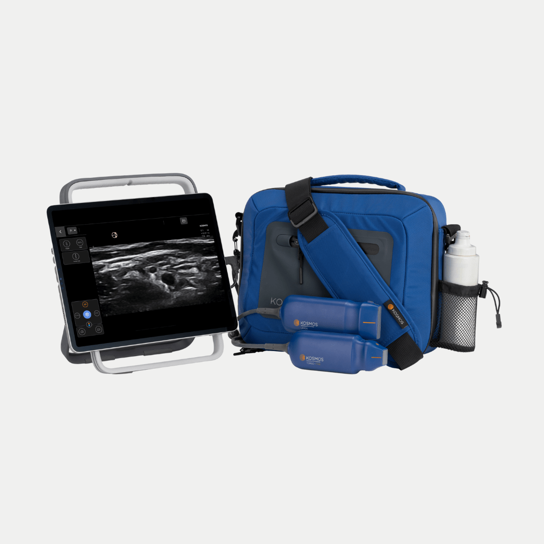
EchoNous, with its Kosmos system, leads the way in innovation by integrating proven Piezo technology with advanced imaging capabilities like dynamic beam forming and noise reduction. This article explores the mechanics, physics, and types of ultrasound probes while articulating why EchoNous remains a trusted leader in imaging technology.
Ready to enhance your diagnostic accuracy? Explore Kosmos by EchoNous today!
Key Takeaways
- POCUS relies on ultrasound probes to convert energy into diagnostic images
- Piezoelectric crystals are the gold standard for reliable, high-resolution imaging
- CMUT and PMUT are emerging alternatives but face performance challenges
- Key physical principles such as noise, frequency, attenuation, and beam forming govern imaging quality
The EchoNous Kosmos system delivers superior precision, adaptability, and reliability
What is an Ultrasound Probe?
The Role of the Probe in POCUS Systems
The ultrasound probe, or transducer, is the linchpin of any POCUS system, functioning as both the sender and receiver of sound waves. This device houses specialized components that convert electrical signals into high-frequency sound waves, which are emitted into the body. These waves interact with tissues, organs, and fluid-filled spaces, bouncing back as echoes that the probe captures and transmits to the ultrasound machine. The machine then processes these signals into real-time images displayed on a screen (3,4).
Ultrasound systems are engineered to deliver precise and consistent imaging, making them indispensable for clinical decision-making. Their ability to visualize internal structures without radiation or the need for invasive procedures has made POCUS a preferred diagnostic tool in a variety of settings, including emergency departments, intensive care units, and primary care offices (5).
The versatility of ultrasound machines extends to their applications, ranging from superficial imaging of tendons and vessels to deep exploration of the heart and abdomen. By enabling real-time imaging, POCUS devices empower clinicians to make rapid, informed decisions at the point of care. This immediacy is particularly valuable in critical situations where every second counts.
In POCUS systems, the ultrasound probe is not merely a diagnostic tool—it’s a bridge between the clinician’s expertise and the patient’s needs, facilitating better outcomes and faster care (6).
Anatomy of an Ultrasound Probe
Ultrasound probes are complex devices meticulously engineered to emit and receive sound waves with precision. Their functionality depends on several key components, each playing a vital role in producing high-quality diagnostic images.
1. Piezoelectric Crystals
At the heart of the probe are piezoelectric Crystals, which generate and detect sound waves. When an electrical current is applied, these crystals vibrate, emitting high-frequency sound waves. Upon receiving echoes from tissue interfaces, the crystals vibrate again, converting mechanical energy into electrical signals for image processing. Their reliability, accuracy and efficiency make them the gold standard for ultrasound imaging.
2. Acoustic lens
The acoustic lens sits on the probe’s surface, shaping and focusing the emitted sound waves into a narrow beam. This focus enhances resolution by ensuring sound waves converge accurately on the target area, reducing noise and improving image clarity.
3. Backing Material
Located behind the Piezo crystals, the backing material absorbs excess vibrations, preventing prolonged ringing of the crystals. This damping effect sharpens the pulses, improving axial resolution and image quality.
4. Electronics and Thermal Management
Some ultrasound systems keep their processing electronics in the laptop / display portion of the system, while others, such as Kosmos, house the entire electronic system within the probe itself. This is essentially a specialized computer that drives the signals to the ultrasound array and processes the imaging coming back from the body. All that computer work can generate significant heat, leading to the need for specialized materials and architecture to dissipate that heat to the outside world.
5. Housing
The external housing protects the probe’s internal components while providing an ergonomic grip for the clinician. It ensures durability and facilitates ease of use, even during extended procedures.
Each of these components works in harmony, enabling ultrasound probes to deliver detailed, reliable images that are critical for effective patient care.
Types of Ultrasound Probes (7)
Linear Probes: For Superficial Structures
Linear probes are designed for high-resolution imaging of shallow structures, making them a staple in Point-of-Care Ultrasound (POCUS) applications. Operating within a high-frequency range of 6-15 MHz, these probes excel in scenarios where detailed visualization of superficial anatomy is required.
Their unique design features a flat, rectangular array of piezoelectric crystals that emit and receive sound waves in parallel lines. This configuration produces a rectangular field of view, ideal for precision imaging.
Linear probes are particularly useful in:
- Vascular imaging: Assessing blood flow, visualizing veins and arteries, and guiding intravenous (IV) access for procedures like PICC line placement or insertion of peripheral IVs
- Musculoskeletal imaging: Evaluating tendons, ligaments, and joint spaces for injuries or abnormalities
- Soft tissue imaging: Detecting superficial masses or fluid collections
The high frequency of linear probes ensures exceptional resolution, capturing fine details of tissues close to the skin surface. However, their depth penetration is limited, making them less suitable for imaging deeper structures beyond 10cm.
By offering clarity and precision in superficial imaging, linear probes are indispensable tools for clinicians performing detailed assessments or procedural guidance in real-time. EchoNous offers the Kosmos Lexsa linear probe for high-frequency imaging, covering applications in Vascular, Lung, Nerve and MSK.
Convex Probes: Wide-Field Imaging
Convex probes, also known as curved array probes, are tools designed for imaging deeper structures within the body. They operate at a lower frequency range of 2-5 MHz, allowing for greater tissue penetration compared to high-frequency probes.
The distinguishing feature of convex probes is their curved array of piezoelectric crystals, which emit sound waves in a fan-shaped pattern. This curved arrangement expands the field of view, enabling clinicians to visualize a larger area in a single scan.
Convex probes are useful in:
- Abdominal imaging: Proving detailed assessments of organs such as the liver, kidneys, and pancreas
- Obstetrics and gynecology: Monitoring fetal development and evaluating pelvic structures
While convex probes sacrifice some resolution compared to linear probes, their ability to penetrate deeper tissues makes them ideal for applications requiring broad, comprehensive views. This balance of depth and field of view makes convex probes helpful for imaging in general medicine and specialty care.
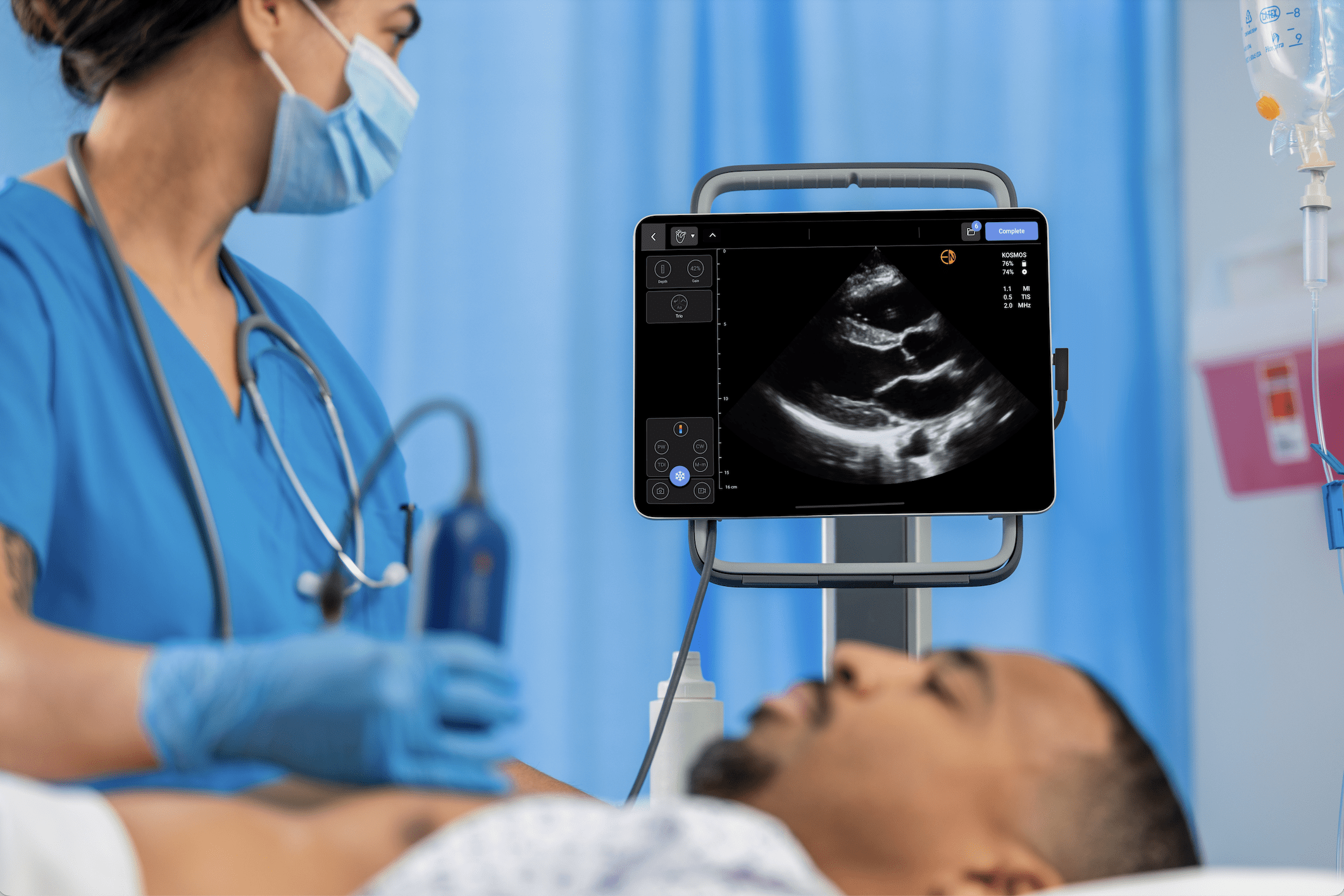
Phased Array Probes: Cardiac Focus
Phased array probes are specifically designed for imaging deep structures in tight anatomical spaces, making them ideal for cardiac, lung, and abdomen imaging as well as other applications like Bladder and Obstetrics, where precision and maneuverability are critical. These probes operate at a frequency range of 2-7 MHz, enabling sufficient penetration to visualize the heart and other internal organs.
The compact, square-shaped design of the phased array scan head allows them to be positioned easily between ribs or in other tight spaces. This is critical to obtaining a good ultrasound “window” to perform an exam. The piezoelectric crystals in phased array probes are arranged in a small, tightly packed configuration and are activated in sequences or “phases” to steer and focus the ultrasound beam dynamically. This advanced beam control ensures clear visualization of moving structures like heart valves and blood flow patterns.
Phased array probes are primarily used for:
- Echocardiography: Assessing cardiac function, valve abnormalities, and structural defects
- Emergency diagnostics: Rapid assessment of cardiac and lung activity and abdominal fluid collection in trauma or critical care settings
- Abdominal exams covering applications in Obstetrics, Gynecology, Family Medicine, and Bladder Volume scanning
In addition to linear, convex, and phased array probes, specialty probes like endocavitary, 3D/4D, and TEE exist to cater to complex scenarios where conventional transducers might fall short.
Looking for a versatile imaging solution? Contact EchoNous to find the right probe for your needs.
The Science Behind Ultrasound Crystals
Piezoelectric Crystals: The Gold Standard
At the core of most ultrasound probes are piezoelectric crystals, the cornerstone technology driving accurate and efficient imaging. These crystals are unique materials that exhibit the piezoelectric effect, a phenomenon where they convert electrical energy into mechanical sound waves and vice versa.
How Piezoelectric Crystals Work
- Electrical Stimulation: When an electric current passes through the crystals, they vibrate, generating high-frequency sound waves.
- Wave Transmission: These sound waves travel into the body, interacting with tissues and reflecting back as echoes when they encounter different densities.
- Echo Reception: Upon receiving the echoes, the crystal vibrates again, converting the returning sound waves into electrical signals.
- Image Formation: The ultrasound machine processes these signals to create detailed diagnostic images.
Why Piezoelectric Crystals Are Preferred
- Proven Reliability: Decades of clinical use have demonstrated their durability and accuracy
- High Resolution: They produce precise, detailed images, which are particularly critical for applications requiring the fine differentiation of tissue layers
- Broad versatility: Piezoelectric crystals support a wide range of frequencies, allowing deep imaging
- Energy Efficiency: These crystals minimize energy loss, ensuring consistent imaging quality
Experience the unmatched precision of piezoelectric technology. Explore Kosmos today!
CMUT: Capacitive Micromachined Ultrasonic Transducers (8,9)
How CMUT Works
- Structure: CMUTs consist of a thin silicon membrane suspended over a cavity, with electrodes on either side.
- Wave Emission: When a voltage is applied, the membrane vibrates, producing ultrasonic waves.
- Wave Reception: Returning echoes cause the membrane to oscillate, generating electrical signals processed into images.
Limitations of CMUT technology:
- Less energy-efficient than piezoelectric crystals, leading to reduced signal strength and image quality, especially at depth
- Prone to failure over relatively short periods of time. CMUT technology cannot match the reliability and resolution of piezo-based systems
PMUT: Piezoelectric Micromachined Ultrasonic Transducers
PMUTs use thin-film piezoelectric materials deposited onto a silicon membrane to create sound waves.
Challenges Compared to Piezoelectric Crystals
- They struggle to achieve the same level of resolution and depth penetration as traditional piezoelectric crystals, limiting their use in applications that require high precision.
- Limited reliability: less robust, affecting longevity
- Not as clinically validated or widely adopted as piezoelectric crystals
Ultrasound Physics Essentials (10,11)
Frequency and Depth Penetration
In ultrasound Imaging, frequency is pivotal in balancing resolution and depth penetration, two key factors for diagnostic clarity. Some companies claim one single probe is sufficient for multiple exam types, but due to physical and frequency limitations, it is difficult to obtain good diagnostic quality. EchoNous offers different transducers to address this issue and allow clinicians to switch between frequencies, ensuring high-resolution imaging for superficial targets while retaining the ability to explore deeper anatomical structures.
High Frequency (6-15 MHz):
- Produces short wavelengths that provide excellent resolution
- Limited depth penetration only, ideal for superficial structures, such as tendons, blood vessels, and skin
Low Frequency (2-5 MHz):
- Generates longer wavelengths, enabling sound waves to penetrate deeper into the body
- Commonly used for abdominal imaging, obstetric scans, and cardiac diagnostics
- Sacrifices fine resolution at shallow depths, leading to less detailed images of superficial structures
Attenuation and Image Clarity
Attenuation refers to the gradual weakening of ultrasound waves as they travel through tissues. This phenomenon reduces wave intensity, impacting the quality of the returning echoes and, consequently, the image clarity.
Attenuation occurs due to:
- Absorption: Conversion of sound wave energy into heat as it travels.
- Scattering: Deflection of sound waves in multiple directions when interacting with heterogeneous tissues.
- Reflection: Partial redirection of waves at tissue boundaries.
To counteract attenuation, ultrasound systems incorporate gain adjustments, which amplify the returning echoes. Key types of gain control include:
- Overall Gain: Adjusts the brightness of the entire image.
- Time Gain Compensation (TGC): Enhances brightness at specific depths, ensuring uniform clarity across the field of view.
By optimizing gain settings, clinicians can maintain diagnostic accuracy, even when imaging deeper structures or encountering tissues with high attenuation properties. EchoNous’ systems excel in managing attenuation through precise gain controls, delivering consistently clear images.
Beamforming and Image Precision
Beamforming is a crucial technique that shapes and focuses ultrasound waves to enhance image resolution and accuracy. It involves controlling the emission and reception of sound waves, ensuring they converge at the target area to create a well-defined image. Dynamic beamforming is now the conventional approach so only two approaches are used today.
Types of Beamforming
- Dynamic Beamforming
- Uses advanced algorithms to continuously adjust the focus as waves travel, improving resolution at varying depths
- Reduces artifacts and noise, resulting in sharper images
- Adaptive Beamforming
- Employs real-time adjustments based on tissue characteristics, optimizing clarity in challenging imaging conditions
EchoNous’ Innovations in Beamforming
EchoNous’ Kosmos system integrates state-of-the-art beam-forming technology to deliver unmatched imaging precision. Features include:
- Dynamic Focus: Ensures consistent resolution across the entire image
- Noise Reduction Algorithms: Minimize interference and enhance quality
- Broadband Beamforming: Combines low and high frequencies to optimize depth and resolution
Through innovative beam-forming techniques, EchoNous empowers clinicians to achieve detailed, accurate diagnostics across various applications. Beamforming is not just a technical process; it is the foundation of superior ultrasound imaging.
Artifacts in Ultrasound Imaging (10,12)
Shadowing
- Occurs when sound waves encounter highly reflective or absorptive structures, such as bones or stones
- Results in a dark, hypoechoic area behind the structure where sound cannot penetrate
Posterior Enhancement
- Seen when sound waves pass through fluid-filled structures like cysts or the bladder
- Fluid allows sound to travel with minimal attenuation, causing tissues behind the fluid to appear brighter than surrounding areas
Reverberation
- Caused by repeated bouncing of sound waves between two highly reflective surfaces
- Appears as multiple parallel lines on the image, often seen near air-filled structures like lungs or bowel loops
Recognizing and Mitigating Artifacts
How to identify Artifacts
- Shadowing: look for consistent dark areas behind dense structures, such as bones or calculi
- Posterior Enhancement: identify abnormality bright areas behind fluid-filled structures
- Reverberation: Recognize repeating, evenly spaced lines that often occur near air-filled or metallic objects
Strategies to Mitigate Artifacts
- Adjust Probe angle: Changing the angle of incidence can reduce reflections and improve visualization of underlying structures
- Optimize Gain Settings: Use appropriate overall gain and Time Gain Compensation (TGC) to balance brightness across the image
- Use Alternative Views: Scanning from different angles or using a different probe type can clarify ambiguous findings
- Leverage Advanced Software: Modern systems like EchoNous’ Kosmos incorporate artifact-reduction algorithms to enhance image clarity
Upgrade your diagnostic capabilities. Request a demo and see Kosmos in action.
Why EchoNous Leads in Ultrasound Technology
Advanced Beamforming Technology
Beamforming shapes and focuses ultrasound waves on optimizing their resolution and accuracy. EchoNous has this cutting-edge beam-forming technology to deliver unparalleled image clarity, sharpness, and artifact-free images for diagnostic precision.
Proven Piezoelectric Crystal Innovation
Accuracy: Piezoelectric Crystals in Kosmos probes are engineered to deliver exceptional energy efficiency, reducing signal loss for stronger and clearer echoes for both superficial and deep structures.
Reliability: Durable and reliable robust piezoelectric technology helps clinicians achieve consistent results, even in high-demand clinical environments. This is why EchoNous transducers can withstand drop tests and we can confidently offer a 5-year warranty on our transducers
Versatility Across Clinical Applications
Unparalleled versatility for a wide range of diagnostic scenarios:
- Cardiac Imaging
- Abdominal Scans
- Vascular studies
- Musculoskeletal and Soft Tissue Diagnostics
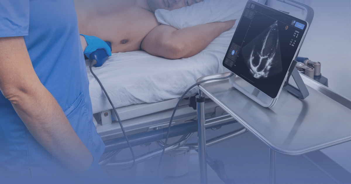
With its portable design, advanced imaging features, and adaptable probes, Kosmos ensures clinicians are equipped to provide accurate diagnostics in any setting-from bedside evaluations to complex clinical environments.
Designed and Built In-house
EchoNous transducers are all designed and made in-house, unlike other companies which often buy transducers off the shelf and re-brand them. Manufacturing in the United States allows EchoNous to maintain higher quality standards and precise craftsmanship, ensuring our products consistently meet superior levels of excellence. In addition, EchoNous US-based service and support teams offer exceptional customer service and same or next-day turnaround for many issues.
Ready to revolutionize your diagnostic capabilities? Explore EchoNous Kosmos and experience the future of ultrasound technology today.
Conclusion
Ultrasound probes and the ultrasound crystals within them are the cornerstone of modern diagnostic imaging. From the trusted reliability of piezoelectric crystals to emerging technologies like CMUT and PMUT, these components enable real-time, non-invasive visualization of the human body. They are critical tools for clinicians, offering detailed images that inform rapid and accurate decisions.
Among these advancements, EchoNous’ Kosmos system stands out as a leader in innovation. By combining proven piezoelectric crystal technology with advanced beamforming and artifact-reduction algorithms, Kosmos delivers unmatched precision and versatility. Its ability to adapt to a wide range of clinical applications – from cardiac and vascular imaging to abdominal and musculoskeletal diagnostics – empowers clinicians to provide superior patient care.
EchoNous’ dedication to integrating state-of-the-art engineering with practical clinical solutions ensures that its technology not only meets but exceeds the demands of today’s medical professionals. The Kosmos system exemplifies how ultrasound innovation can elevate diagnostic imaging, improving outcomes across a variety of care settings.
Take your imaging to the next level. Contact EchoNous to get started today!
References
1. Fraleigh, C. D. M., et al. (2022). Point-of-care ultrasound. The Nurse Practitioner, 47(8), 14–20. PMID: 35877142 PMCID: PMC9301985
2. Cafarelli, A., et al. (2021). Piezoelectric Nanomaterials Activated by Ultrasound: The Pathway from Discovery to Future Clinical Adoption. ACS Nano, 15(7), 11066. PMID: 34251189 PMCID: PMC8397402
3. Ultrasound. (n.d.). National Institute of Biomedical Imaging and Bioengineering.
4. Borowy, C. S., et al. (2024). Sonography Physical Principles And Instrumentation. In StatPearls. StatPearls Publishing.
5. Magon, F., et al. (2024). Point-of-Care Ultrasound (POCUS) in Adult Cardiac Arrest: Clinical Review. Diagnostics, 14(4), Article 4.
6. Andersen, C. A., et al. (2019). Point-of-Care Ultrasound in General Practice: A Systematic Review. The Annals of Family Medicine, 17(1), 61–69. PMID: 30670398 PMCID: PMC6342599
7. BGill. (2019, March 27). Ultrasound Probes | The Break Down. Probo Medical.
8. Salim, M. S., et al. (2012). Capacitive Micromachined Ultrasonic Transducers: Technology and Application. Journal of Medical Ultrasound, 20(1), 8–31.
9. Wang, J., et al. (2020). Capacitive micromachined ultrasound transducers for intravascular ultrasound imaging. Microsystems & Nanoengineering, 6(1), 1–13. PMID: 34567683 PMCID: PMC8433336
10. Grogan, S. P., et al. (2024). Ultrasound Physics and Instrumentation. In StatPearls. StatPearls Publishing.
11. Ultrasound Physics and Technical Facts for the Beginner. (2020, July).12. Kremkau, F. W., et al. (1986).
12. Artifacts in ultrasound imaging.Journal of Ultrasound in Medicine: Official Journal of the American Institute of Ultrasound in Medicine, 5(4), 227–237.