Mastering Musculoskeletal (MSK) Ultrasound
Applications, Techniques and Benefits
Musculoskeletal ultrasound (MSK ultrasound) is an invaluable diagnostic and therapeutic tool utilized across many medical specialties. This guide introduces the fundamentals of musculoskeletal ultrasound, its wide-ranging applications, and the benefits of MSK ultrasound. Understanding these aspects is essential for healthcare professionals aiming to enhance their diagnostic capabilities and improve patient outcomes.
Basics of Musculoskeletal Ultrasound
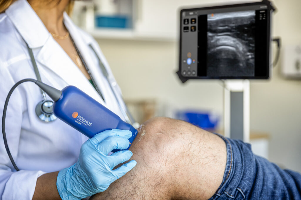
Musculoskeletal ultrasound is a specialized imaging technique used to assess and diagnose disorders related to muscles, tendons, ligaments, joints, and soft tissues. The underlying principle involves the use of high-frequency sound waves that are emitted by a transducer, penetrate the body, and reflect off tissues to create real-time images. This technology is particularly suited for imaging superficial structures with high resolution.
The ultrasound system comprises a machine with a monitor, various types of probes (or transducers), and specific settings tailored for optimal imaging of musculoskeletal structures. Probes commonly used include linear array transducers, which provide high-resolution images ideal for evaluating tendons and ligaments.
Applications of Musculoskeletal Ultrasound
Musculoskeletal ultrasound serves a broad range of diagnostic purposes. It is highly effective in diagnosing tendon tears, muscle injuries, ligament sprains, and joint abnormalities. For instance, it can reveal partial or complete tendon tears in the shoulder, muscle strains in the leg, or ligament sprains in the ankle.
In addition to diagnosis, MSK ultrasound is instrumental in guiding various interventional procedures. It assists in the precise administration of injections, aspirations, and biopsies by providing real-time visualization of the target area. This guidance enhances the accuracy and safety of such procedures.
The technology is also valuable for monitoring chronic conditions and disease progression. In cases of rheumatoid arthritis or osteoarthritis, MSK ultrasound can track changes in joint and tendon structure over time, aiding in the management and adjustment of treatment plans.
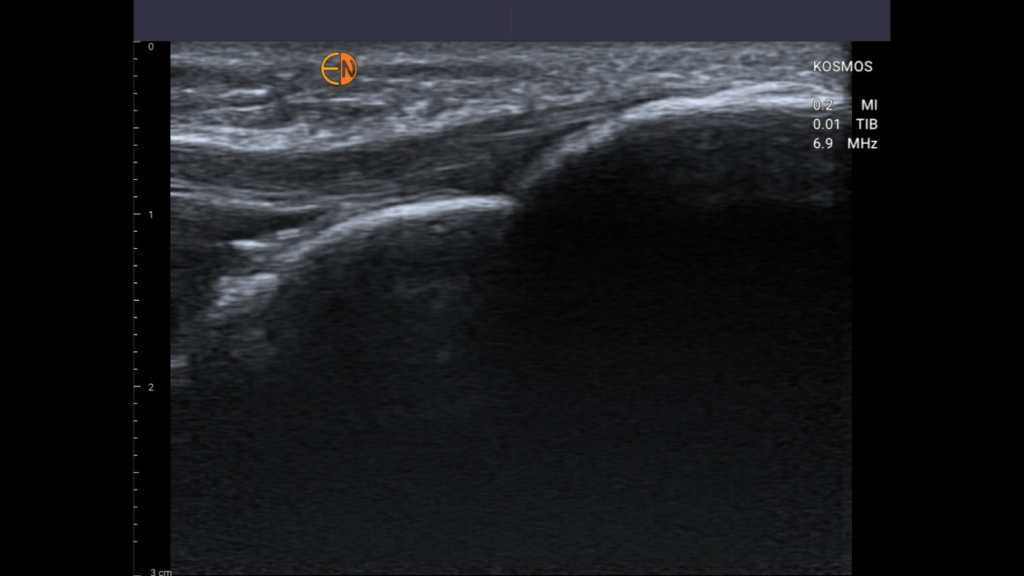
Tibial Insertion of Patella Tendon
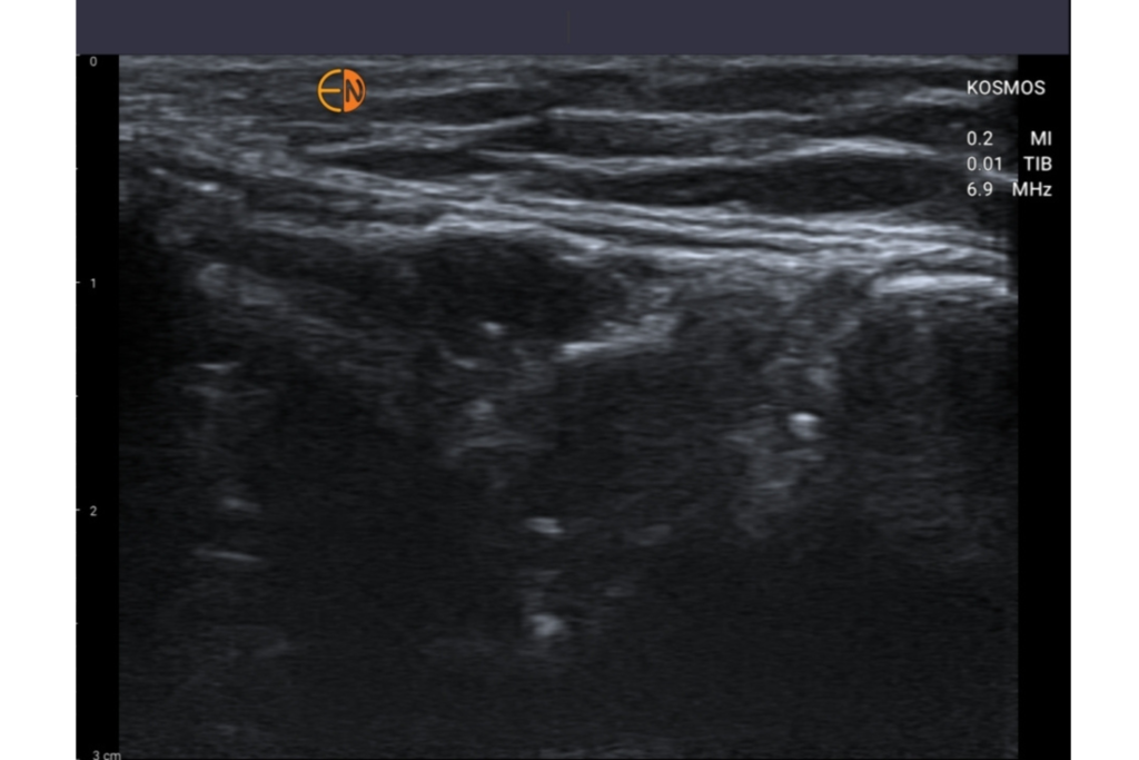
Medial Collateral Ligament and Pathology
Benefits of Musculoskeletal Ultrasound
One of the primary advantages of musculoskeletal ultrasound is its safety and non-invasiveness. Unlike X-rays or CT scans, ultrasound does not use ionizing radiation, making it a safer option, especially for repeated use.
Another significant benefit is the ability to perform real-time imaging, which allows dynamic assessment of musculoskeletal structures during movement. This capability is particularly useful for diagnosing conditions that only manifest during certain motions or activities.
Musculoskeletal ultrasound is also highly accessible and cost-effective compared to other imaging modalities like MRI. It is relatively inexpensive and widely available, making it a practical choice for many healthcare settings.
Tenchiques in Musculoskeletal Ultrasound
Effective use of musculoskeletal ultrasound relies on proper scanning techniques and protocols. Standard scanning protocols vary for different parts of the body, such as the shoulder, knee, and ankle, each requiring specific positioning and preparation to obtain the best diagnostic images.
Patient preparation involves ensuring comfort and proper positioning to expose the area of interest. Image optimization is crucial, requiring adjustments to the ultrasound settings to enhance image clarity and accuracy. Techniques such as adjusting the frequency, gain, and focus of the ultrasound beam can significantly improve the quality of the images obtained.
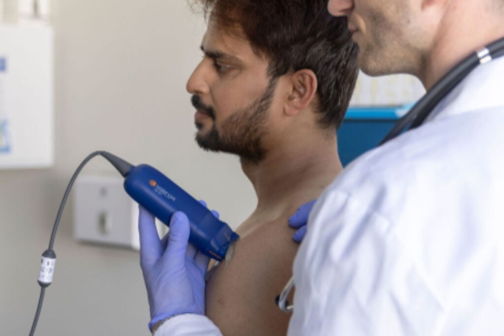
Challenges and Limitations
Despite its many benefits, musculoskeletal ultrasound has certain limitations. The effectiveness of the imaging is highly dependent on the skill of the operator, making training and experience essential for accurate diagnosis. Imaging depth can also be a challenge, particularly in patients with a high body mass index, where deeper structures may be more difficult to visualize.
When compared to MRI, ultrasound may have limitations in image resolution and depth of imaging capabilities, which can impact the ability to diagnose certain conditions. However, advancements in ultrasound technology continue to mitigate these limitations.
Advances and Future Directions
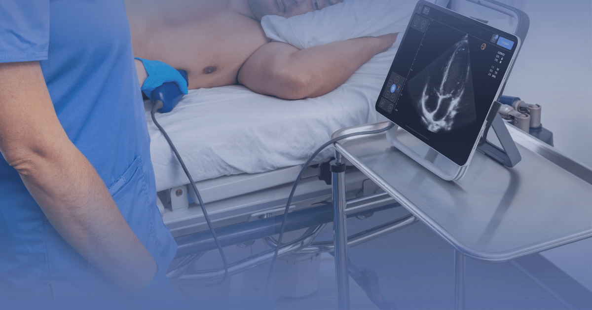
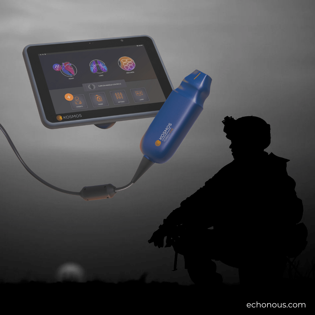
Recent technological advancements have significantly enhanced the capabilities of musculoskeletal ultrasound. Improvements in image clarity, the development of portable devices, and the integration of wireless technology are some of the notable innovations. These advancements expand the utility and convenience of ultrasound in various clinical settings.
Looking ahead, new therapeutic applications and potential integration with other imaging technologies are on the horizon. These developments promise to further enhance the diagnostic and therapeutic potential of musculoskeletal ultrasound.
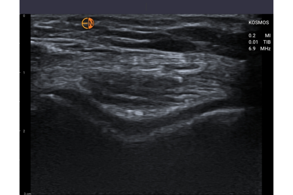
Articular Cartilage of the Trochlea
Training and Education
For healthcare professionals interested in specializing in musculoskeletal ultrasound, various learning pathways and training requirements must be met. Comprehensive training programs and continuing education are crucial for mastering the techniques and staying updated with the latest advancements. Ongoing education ensures that practitioners maintain their skills and provide the highest quality of care.
Interested in using POCUS for MSK APplications?
Musculoskeletal ultrasound is a powerful tool in modern medicine, offering significant diagnostic and therapeutic potential. Its safety, accessibility, and real-time imaging capabilities make it an invaluable asset in various medical specialties. Healthcare professionals are encouraged to consider training in musculoskeletal ultrasound to enhance their diagnostic capabilities and improve patient outcomes.
We invite healthcare professionals to explore training courses or workshops in musculoskeletal ultrasound. For more detailed advice or to schedule a consultation with an expert, please contact us. Our team is here to support your journey in mastering this essential imaging tool.
