Identifying & Diagnosing Ischemic Mitral Regurgitation
Patient: A 51 year old female patient presented to the ER with gradually worsening shortness of breath.
Initial Examination: She had a history of coronary artery disease and ischemic mitral regurgitation treated with CABG and mitral valve repair three years earlier. On clinical examination a holosystolic apical murmur was noted. Her jugular veins were dilated and lower limb edema was present.
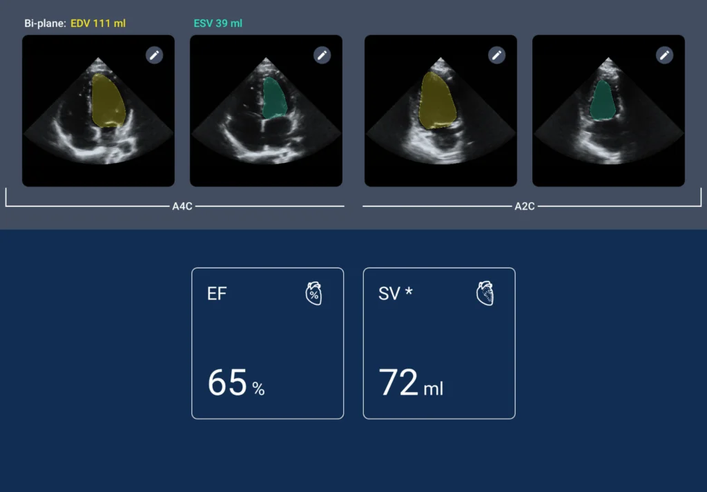
Kosmos POCUS Examination: Bedside examination with KOSMOS revealed impaired systolic function of both ventricles accompanied by severe residual ischemic mitral regurgitation and significant functional tricuspid regurgitation. Intravenous diuresis was promptly initiated resulting in improvement of her symptoms.
PLAX view with color showing severe residual mitral regurgitation
RV inflow view
RV inflow view with color showing significant tricuspid regurgitation
CW TR signal from RV inflow view
PSAX view at the level of the papillary muscles demonstrating severely impaired LV systolic function and the presence of segmental wall motion abnormalities
A4C view showing dilated LV with segmental wall motion abnormalities and impaired LV systolic function. The RV is hyperdynamic with overall mildly impaired RV systolic function. There is lack of coaptation of the TV leaflets. Bi-atrial enlargement is also evident.
A4C view with color on the TV showing severe TR
CW TR signal from the A4C view
Assessment of TAPSE with the use of M-mode from the A4C view
A4C view with color on the repaired mitral valve showing severe residual MR
CW MR signal from the A4C view
Subcostal view showing dilated IVC with minimal respiratory variation
PW Doppler in the hepatic veins demonstrating systolic flow reversal suggestive of severe TR


