How Do You Perform a DVT Ultrasound? The Two-Point Compression Exam
Is that swollen leg a clot or something else? The two-point compression exam helps clinicians answer that question at the bedside. A two-point compression exam is a focused deep vein thrombosis (DVT) ultrasound technique used to assess the common femoral and popliteal veins for signs of thrombosis.1 Using direct pressure from the ultrasound transducer, the clinician can see whether the veins compress normally. A vein that does not fully compress can be a sign of proximal DVT.
Key Takeaways:
- The two-point exam compresses the common femoral vein and the popliteal vein. A vein that does not fully collapse under transducer pressure raises concern for DVT.1
- Emergency physician-performed ultrasound for DVT has shown a sensitivity of 96.1% and specificity of 96.8% compared with reference imaging.2
- About 6% of lower-extremity DVTs are isolated outside the common femoral and popliteal veins, so a negative two-point exam does not exclude DVT.1
- Interpret every result alongside pre-test probability. Patients with higher pre-test probability and a negative exam may need additional or serial ultrasound.3
What is DVT?
Deep vein thrombosis occurs when a blood clot forms deep within a vein, most commonly in the lower extremities. A primary concern with a DVT is that the clot can propagate or fully dislodge and become a life-threatening pulmonary embolism. Pulmonary emboli have an estimated mortality rate of 10% to 30%, making it important to recognize and evaluate DVT when suspected.1
When should you use DVT ultrasound?
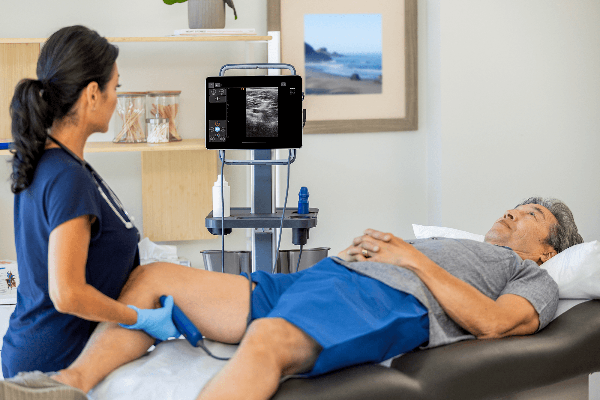
Ultrasound can be used when a patient has signs or symptoms that raise concern for a DVT, such as leg pain or swelling. Leg swelling has many possible causes, including cellulitis, which is why point-of-care ultrasound (POCUS) is useful for narrowing the cause at the bedside. A focused two-point compression ultrasound exam can quickly assess for thrombus in the common femoral and popliteal veins at the bedside.
In critically ill patients, this assessment may also be performed alongside other bedside ultrasound procedures. The results can help determine whether further testing or treatment for suspected DVT is needed, depending on the patient’s overall clinical picture.
There is substantial evidence supporting the use of bedside ultrasound for evaluating suspected DVT. In one systematic review of 16 studies including more than 2,300 patients, emergency physician-performed ultrasound had a sensitivity of 96.1% and specificity of 96.8% compared with reference imaging.2
Table of Contents
- How Do You Perform a DVT Ultrasound? The Two-Point Compression Exam
- What is DVT?
- When should you use DVT ultrasound?
- How to perform a two-point compression DVT ultrasound
- How do you interpret the findings?
- How does pre-test probability change the result?
- Limitations and common pitfalls
- Putting the two-point exam into practice
- Q&A: DVT ultrasound and the two-point compression exam
- References
How to perform a two-point compression DVT ultrasound
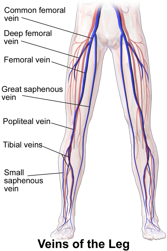
The exam has four steps: select a linear transducer, position the patient, then compress the common femoral vein and the popliteal vein at set points. At each point, the question is whether the vein walls fully come together. Figure 1 shows the deep veins of the leg.
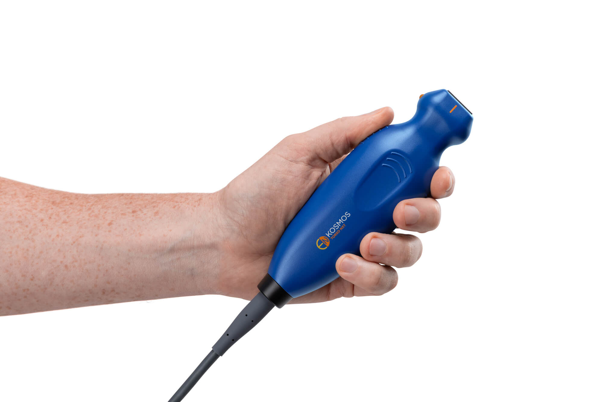
- Select the transducer. Use a high-frequency linear transducer, such as the Kosmos Lexsa probe, with the vascular setting. Orient the transducer with the indicator toward the patient’s right side.
- Position the patient. For the common femoral vein (CFV), position the leg slightly externally rotated with the knee slightly flexed in a “frog-leg” position. For the popliteal vein, keep the knee slightly flexed and position the transducer over the proximal medial popliteal region. The patient can also be positioned prone or in a lateral decubitus position for popliteal imaging, depending on access and comfort.
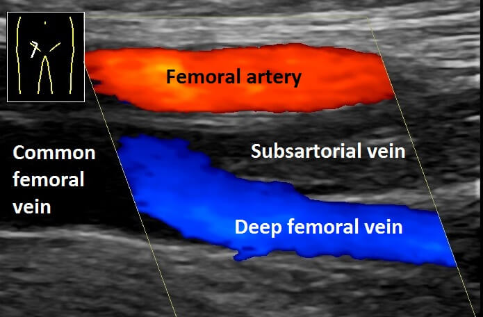
- Evaluate the femoral vein. The CFV runs beneath the inguinal ligament and continues down the inner thigh. The inguinal crease is a useful surface landmark for locating the vein. Start at the inguinal crease with the transducer in a transverse orientation and locate the CFV (Figure 2). If you see three vessels (the common femoral artery, common femoral vein, and saphenous vein), move the transducer more proximal toward the inguinal ligament. Center the vein and apply perpendicular pressure until it completely compresses, using enough pressure to bring the vein walls together while slightly compressing the artery. Move 1 to 2 cm distally to assess the great saphenous vein confluence, then another 1 to 2 cm to assess the deep femoral and femoral vein junction, repeating compression at each location.
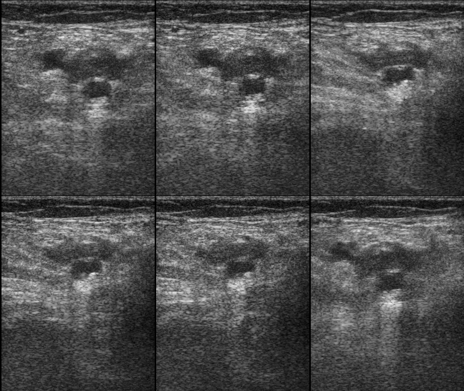
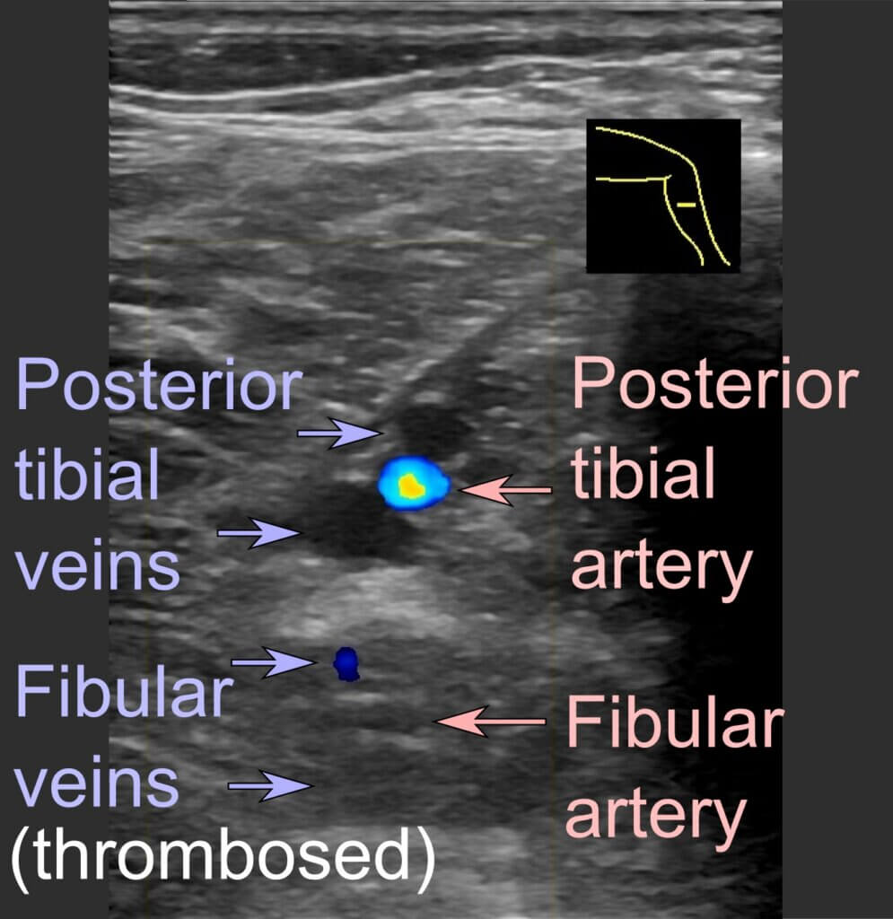
- Evaluate the popliteal vein. Next, move the transducer to the proximal medial popliteal region with the knee slightly flexed. Identify the popliteal vein and artery, center the vein, and apply perpendicular pressure to assess for complete compression (Figure 3). For a more extensive lower-extremity exam, follow the popliteal vein distally toward the trifurcation into the posterior tibial, anterior tibial, and peroneal veins and compress these vessels as well (Figure 4).1
Most importantly, follow a consistent sequence. At each location, identify and center the vein, then apply perpendicular compression until the vein walls come together. A vein that does not fully compress may indicate DVT and should be interpreted along with the patient’s clinical presentation and other exam findings.
How do you interpret the findings?
Normal: The vein completely collapses with transducer pressure. The vein walls should come together with compression, while the adjacent artery remains pulsatile and does not fully collapse.1
Abnormal: The vein does not completely compress despite appropriate pressure. This noncompressibility is the key ultrasound finding that raises concern for DVT. If a thrombus is directly visualized within the vein, that can further support the diagnosis.1
The DVT compression ultrasound is focused on the common femoral and popliteal veins and a negative exam does not exclude thrombus in other portions of the lower extremity.1 The ultrasound findings should therefore be interpreted along with the patient’s symptoms, clinical pre-test probability, and any additional testing that may be indicated.3
Findings should be interpreted by a treating physician with appropriate training in limited compression ultrasound, following specialty-specific or organizational guidelines. Credentialing requirements vary by institution and specialty.4
How does pre-test probability change the result?
Ultrasound findings are only part of the diagnostic picture. The patient’s pre-test probability, or the likelihood of DVT before testing based on their clinical presentation and risk factors, also affects how the ultrasound result should be interpreted. Symptoms such as unilateral leg swelling, localized tenderness, and swelling of the entire leg can raise suspicion for DVT. Risk factors include a history of DVT, active cancer, recent surgery, prolonged immobility, and lower-extremity injury or immobilization. The Wells score estimates this probability based on clinical findings and risk factors. Several versions of the scoring model are used to estimate pre-test probability for DVT, and the approach may vary by institution or clinical guideline. The three-level model below assigns points with scores of 3 or higher indicating high probability.3
| Component | Points |
|---|---|
| Active cancer | 1 |
| Localized tenderness | 1 |
| Entire leg swollen | 1 |
| Calf swelling > 3 cm | 1 |
| Pitting edema | 1 |
| Collateral superficial veins | 1 |
| Previous DVT | 1 |
| Bedridden/surgery | 1 |
| Paralysis | 1 |
| Alternate diagnosis | -2 |
A negative ultrasound does not have the same meaning in every patient. In patients with a lower pre-test probability, a negative study may be enough to help exclude DVT, while patients with a higher pre-test probability may need additional or serial ultrasound testing if the initial study is negative.3
Limitations and common pitfalls
A two-point compression exam focuses on the common femoral and popliteal veins and does not assess the entire deep venous system of the lower extremity. In fact, about 6% of lower-extremity DVTs are isolated outside the common femoral and popliteal veins and may not be detected with a simple two-point exam.1
The technique used during the exam can also affect findings. The transducer should be perpendicular to the vessel, and enough pressure should be applied to bring the vein walls together. Inadequate or oblique compression can make a normal vein appear noncompressible and lead to a false-positive finding.1
Image quality can be affected by patient factors as well. In patients with obesity, a curvilinear transducer may provide better penetration while maintaining enough resolution to evaluate the deep veins.1
Finally, it is important to recognize structures that can mimic DVT. Enlarged lymph nodes, superficial veins, Baker’s cysts, abscesses, and hematomas can appear as noncompressible structures or be mistaken for deep veins. Increasing the depth can help identify the appropriate deep veins, while Baker’s cysts may be distinguished by their larger size and location extending toward the knee joint.1
Putting the two-point exam into practice
The two-point compression exam answers one focused question at the bedside: do the proximal deep veins compress normally? Paired with a structured pre-test probability assessment, the result helps determine whether further testing or treatment for suspected DVT is needed. Its value depends on consistent technique and a clear understanding of what the exam does not cover.
A checklist for each exam:
- Estimate pre-test probability before scanning.
- Use a high-frequency linear transducer with the vascular setting.
- Compress the common femoral vein at the inguinal crease, the great saphenous vein confluence, and the deep femoral and femoral vein junction.
- Compress the popliteal vein in the popliteal fossa.
- Apply perpendicular pressure until the vein walls meet, and confirm full collapse at each point.
- If the exam is negative and pre-test probability is high, plan additional or serial ultrasound per local protocol.
Kosmos can support clinicians doing lower-extremity venous exams with structured reporting on the Lexsa linear probe as part of its vascular capabilities. To see it in your own clinical workflow, schedule a demo with our team.
Q&A: DVT ultrasound and the two-point compression exam
Yes. A history of DVT can make recurrent thrombosis more challenging to evaluate because residual changes from a previous clot can affect the ultrasound appearance. When recurrent DVT is suspected, prior imaging can be useful for comparison when available.3
A negative two-point exam does not completely exclude DVT, particularly when a thrombus may be located outside the common femoral or popliteal veins. Depending on the patient’s clinical risk and diagnostic strategy, additional testing, systematic compression, or follow-up ultrasounds may be appropriate.1,3
No. A two-point exam evaluates the common femoral and popliteal veins, so an isolated calf DVT would not be detected by this exam.1 A more extensive or whole-leg ultrasound may be needed when distal DVT is suspected, depending on the patient’s clinical presentation and diagnostic strategy.3
Compression is the primary assessment in a focused two-point exam because venous noncompressibility is the key finding. Color Doppler or Pulsed Wave Doppler can be used as an adjunct for assessing venous flow, including with augmentation, but they do not replace adequate compression.1 For more information on using POCUS to evaluate peripheral vascular conditions, see peripheral vascular emergencies.
References
- American College of Emergency Physicians. Deep Vein Thrombosis (DVT). ACEP Sonoguide. Accessed October 2, 2026. Available from: https://www.acep.org/sonoguide/basic/dvt
- Pomero F, Dentali F, Borretta V, et al. Accuracy of emergency physician-performed ultrasonography in the diagnosis of deep-vein thrombosis: a systematic review and meta-analysis. Thromb Haemost. 2013;109(1):137-145. doi:10.1160/TH12-07-0473
- American Society of Hematology. VTE Diagnosis: Teaching Slide Set. 2023. Available from: https://www.hematology.org/-/media/hematology/files/clinicians/guidelines/vte/ash-teaching-slide-setvtediagnosispdffinal71923.pdf
- American College of Emergency Physicians. Emergency Ultrasound Imaging Criteria Compendium: Venous Thrombosis. Approved October 2014. Available from: https://www.acep.org/siteassets/uploads/uploaded-files/acep/by-medical-focus/ultrasound/emergency-ultrasound-imaging-criteria-compendium.pdf