Bowel Ultrasound Complete Guide for Family Physicians to Master IBD Care
GIT complaints are very common in family medicine. Bloating, diarrhea, and abdominal pain are the main symptoms indicating the presence of an inflammatory bowel disease such as ulcerative colitis or Crohn’s disease.
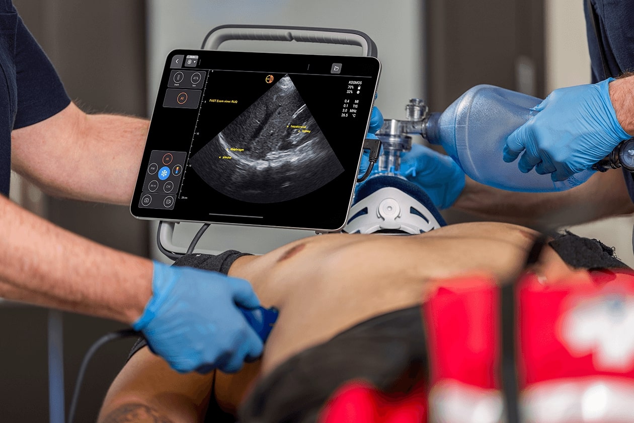
Computed tomography (CT) and magnetic resonance imaging (MRI) are the main tools used to diagnose bowel pathology. Recently, the use of ultrasound has emerged as an effective method to diagnose bowel problems without the radiation, cost, or logistical issues of CT and MRI. Bowel ultrasound, especially POCUS, is a non-invasive tool that can be moved to the bedside to perform the examination and give the same results as CT and MRI.
In IBD, ultrasound offers insights about the wall and even the luminal contents. In this article, we will explore how ultrasound equips family physicians to tackle IBD. It is not a secondary option anymore; it is the front-line management tool.
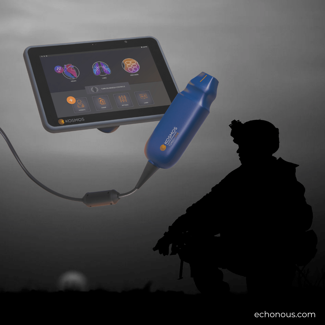
Read more about the Kosmos Advantages in Family Medicine & Primary Care and how the use of ultrasound can enhance your decision-making.
Key Takeaways:
- Bowel Ultrasound: Bowel ultrasound is a cost-effective, accurate, and radiation-free tool. Matching the level of accuracy of CT and MRI in diagnosing IBD conditions. It provides real-time insights into bowel wall and luminal conditions.
- IBD Monitoring: About 30% of IBD patients usually have active inflammation despite being symptomless. Bowel ultrasound is a great tool in regular IBD monitoring and catching what clinicians might miss by taking history.
- Transperineal Ultrasound: Transperineal ultrasound matches the accuracy of MRI in mapping perianal fistulas in IBD conditions. It offers bedside accuracy without the need for sedation or delays.
- Intestinal Ultrasound: Intestinal ultrasound gives real-time insights into luminal contents, such as fluids in intestinal obstruction cases and phytobezoars in Crohn’s. This helps family physicians in broadening diagnostic reach beyond other imaging modalities focusing on the intestinal walls.
Table of contents
- Bowel Ultrasound Complete Guide for Family Physicians to Master IBD Care
Why Bowel Ultrasound Is Important in Family Medicine?
IBD is a common disease seen by family medicine clinicians. It usually affects young adults suffering from long-term flares of remission and flares. Because it has an unpredictable nature, frequent monitoring is necessary because depending on symptoms and clinical correlation alone can be misleading. According to studies, about 30% of IBD patients suffer from active inflammation even though they feel well. (1)
Therefore, depending on symptoms and complaints may lead to missing the mark. Bowel ultrasound acts like a magic tool for IBD patients. It is radiation-free, cost-effective, and can give family physicians valuable, real-time insights during a routine visit. Studies show that using ultrasound along with CT and MRI for IBD can enhance the specificity and sensitivity by more than 90%, with the possibility of using ultrasound stretches even beyond IBD. (2)
For example, an intestinal ultrasound can help in detecting luminal leaks, fluid in small bowel obstruction, and fecal material inside the bowel in constipation. (3)
This broadens the diagnostic reach, which means quicker diagnoses, fewer specialist referrals, and patients not exposed to radiation or additional costs. After all, family medicine is all about time and trust, and bowel ultrasound empowers physicians to enhance them.
Bowel Ultrasound: The Science Behind It
Once you start gliding the probe across the abdomen, ultrasound will help you to identify the five layers of the gut – mucosa, muscularis mucosa, submucosa, muscularis propria, and serosa – in detail. Normal wall thickness is less than 3 mm, while the colon is less than 5 mm if not distended. If the wall is thicker than these measurements, there is definitely a pathological problem. Here are some insights into what intestinal ultrasound can provide:
- Intestinal ultrasound can actually compete with CT and MRI in determining mural inflammation, strictures, and fistulas. (4)
- Color doppler imaging can show hypervascularity, indicating hyperaemia in active Crohn’s.
- Contrast-enhanced ultrasound (CEUS) can quantify mural perfusion. A peak enhancement (PE) decline from 25 dB to 15 dB signals waning inflammation. (5)
- Shear wave elastography (SWE) can match MRI’s precision in measuring stiffness (values over 2 m/s suggest fibrotic strictures).
Intestinal Ultrasound for Assessing Luminal Content
A bowel ultrasound is the perfect tool to assess luminal content.
- In the stomach, clear fluids are anechoic with a “starry night” appearance due to the presence of gas bubbles. On the other hand, solid contents show mixed echogenicity, which is crucial for estimating gastric emptying.
- If the small bowel loops contain excessive fluid and gas, it may indicate that there is a bacterial overgrowth.
- In Crohn’s disease, a stricture might trap a phytobezoar, which is usually visible on ultrasound and not on CT scans.
All these insights make ultrasound a versatile tool, competing with CT imaging’s depth and real-time insights.
Ultrasound vs. CT and MRI in Bowel Ultrasound
If you are still skeptical that ultrasound can provide you with the same results as CT and MRI, here is the evidence.
- Studies show that bowel ultrasound is as accurate as CT and MRI in IBD since it can provide information about wall thickening, abscesses, and fistulas. (6)
- In acute cases, ultrasound can detect diverticulitis, appendicitis, and other acute abdomen cases as reliably as CT. (7)
- Ultrasound can detect small bowel obstruction with an accuracy level of 81.3%. This percentage increases to 91.7% without excess gas. It pinpoints dilated, fluid-filled loops and transition points. CT is the first choice in these cases, but ultrasound is faster and safer. Besides, CT and MRI are much more expensive and include radiation exposure.
- In IBD monitoring, CEUS provides detailed insights about inflammation with the same precision as MRI, which saves the patient the contrast risks.
Transperineal Ultrasound: A Special Technique for Deeper Insights
Transperineal Ultrasound is one of the most powerful techniques for mapping perianal fistulas in IBD. It matches MRI in the level of accuracy since it can reveal hypoechoic tracts and abscesses in the anal canal. (8)
This can guide immediate therapy without delay or the need for sedation. In addition, endovaginal ultrasound can uncover pelvic strictures and inflammation, like in Crohn’s, where the terminal ileum may be missed transabdominally.
Specialized techniques like transperineal for detecting fistulas, endovaginal for pelvic problems, and bowel ultrasound for obstructions make POCUS a bedside match for CT and MRI. It perfectly suits family physicians.
How to Integrate Bowel Ultrasound into Routine Family Practice?
Using bowel ultrasound in family practice is not just about cost or capabilities but also about integrating it in the day-to-day workflows to provide the level of diagnostic accuracy as CT and MRI. Practical protocols include:
- A 6-hour fast to optimize views
- Low-frequency probes will help to survey the abdomen, while high-frequency probes will help zoom into important areas such as the terminal ileum.
- For luminal checks, to avoid common IBD similar conditions like constipation and obstructions, a quick scan should be performed to address the issues with certain measurements in mind. For example, bowel loops are considered dilated if they are over 25mm, and rectal diameter over 0.189 ratio means that there is faecal impaction.
If you are a family physician, you probably had a 35-year-old suffering from bloating after an IBD flare. After performing an intestinal ultrasound, you found fluid-filled jejunal loops, detecting that there might be SIBO. After a breath test, the diagnosis is confirmed. All of this can be done in practice. Of course, training matters. (9)
Beginners should start with acute pain conditions such as appendicitis and diverticulitis, where ultrasound accuracy is high and the conditions are obvious. After that, you can scale to chronic conditions. Most family practices now have portable ultrasound machines, and CEUS/SWE upgrades are affordable.
For patients, this is far more convenient than other imaging modalities since there is no IV contrast, no delays, and fewer visits to the hospital. Besides, you can discuss the results immediately after the scan with the patient without waiting for the report. (10)
Frequently Asked Questions about Bowel Ultrasound
- How Accurate is Bowel Ultrasound compared to MRI and CT for IBD diagnosis?
Bowel ultrasound matches the accuracy of MRI and CT in IBD diagnosis when done right. It can detect IBD hallmarks such as fistulas and wall thickening. For obstructions, the level of accuracy is between 81.3% to 91.7% depending on the amount of gases in the bowel. (11)
- Can Transperineal Ultrasound Replace MRI for Fistula’s Diagnosis?
Yes, it can. While MRI is the gold standard for mapping perianal fistulas in IBD, transperineal ultrasound offers bedside accuracy without the need for sedation.
- What Training Do Family Physicians Need to Perform Bowel Ultrasound?
First, you need the basic POCUS skills such as probe handling and imaging interpretations. After that, you can scale up and start diving deeper into ultrasound.Read more about the impact of POCUS in family medicine.
Conclusion
Bowel ultrasound and POCUS are diagnostic tools equalling the accuracy of CT and MRI. It even surpasses them in cost, speed, and safety. Intestinal ultrasound can help in tracking inflammation and contents, transperineal ultrasound can map fistulas, and CEUS/SWE enhances precision. All of this can be done in 15 minutes in a family practice at the bedside.
Looking to add POCUS capabilities to your practice? Request a Demo today.
References
- Dolinger, M. T., & Kayal, M. (2023). Intestinal ultrasound as a non-invasive tool to monitor inflammatory bowel disease activity and guide clinical decision making. World journal of gastroenterology, 29(15), 2272–2282. https://doi.org/10.3748/wjg.v29.i15.2272
- Fraquelli, M., Colli, A., Casazza, G., Paggi, S., Colucci, A., Massironi, S., Duca, P., & Conte, D. (2005). Role of US in detection of Crohn disease: meta-analysis. Radiology, 236(1), 95–101. https://doi.org/10.1148/radiol.2361040799
- Su, H. Y., Taylor, K. M., Friedman, A. B., Cataletti, G., & Maconi, G. (2024). Ultrasound assessment of gastrointestinal luminal contents: a narrative review. Journal of ultrasound, 27(4), 781–792. https://doi.org/10.1007/s40477-024-00951-3
- Shankar, N., Kuo, L., Krugliak Cleveland, N., Galen, B., Samel, N. S., Perez-Sanchez, A., Nathanson, R., Coss, E., Echavarria, J., Rubin, D. T., & Soni, N. J. (2025). Point-of-Care Ultrasound in Gastroenterology and Hepatology. Clinical gastroenterology and hepatology : the official clinical practice journal of the American Gastroenterological Association, S1542-3565(25)00019-9. Advance online publication. https://doi.org/10.1016/j.cgh.2024.09.040
- Medellin, A., Merrill, C., & Wilson, S. R. (2018). Role of contrast-enhanced ultrasound in evaluation of the bowel. Abdominal radiology (New York), 43(4), 918–933. https://doi.org/10.1007/s00261-017-1399-6
- Nylund, K., Maconi, G., Hollerweger, A., Ripolles, T., Pallotta, N., Higginson, A., Serra, C., Dietrich, C. F., Sporea, I., Saftoiu, A., Dirks, K., Hausken, T., Calabrese, E., Romanini, L., Maaser, C., Nuernberg, D., & Gilja, O. H. (2017). EFSUMB Recommendations and Guidelines for Gastrointestinal Ultrasound. EFSUMB-Empfehlungen und Leitlinien des Gastrointestinalen Ultraschalls – Teil 1: Untersuchungstechniken und Normalbefund (Kurzversion). Ultraschall in der Medizin (Stuttgart, Germany : 1980), 38(3), 273–284. https://doi.org/10.1055/s-0042-115410
- Lawler F. (1992). Sonography in the diagnosis of acute appendicitis. American family physician, 45(6), 2482–2485.
- Wright, E. K., Novak, K. L., Lu, C., Panaccione, R., Ghosh, S., & Wilson, S. R. (2015). Transperineal ultrasonography in perianal Crohn disease: A valuable imaging modality. Canadian journal of gastroenterology & hepatology, 29(8), 445–447. https://doi.org/10.1155/2015/120123
- Mihai, V. C., Gheorghe, L., Rezuș, I. I., Jucan, A. E., Andronic, M. C., Gavrilescu, O., Dranga, M., Andronic, A. M., Prelipcean, C. C., Rezuș, C., & Mihai, C. (2024). Novelties and Perspectives of Intestinal Ultrasound in the Personalised Management of Patients with Inflammatory Bowel Diseases-A Systematic Review. Diagnostics (Basel, Switzerland), 14(8), 812. https://doi.org/10.3390/diagnostics14080812
- Medellin, A., & Wilson, S. R. (2025). Bowel Ultrasound. Radiologic clinics of North America, 63(1), 83–96. https://doi.org/10.1016/j.rcl.2024.08.002
- Su, H. Y., Taylor, K. M., Friedman, A. B., Cataletti, G., & Maconi, G. (2024). Ultrasound assessment of gastrointestinal luminal contents: a narrative review. Journal of ultrasound, 27(4), 781–792. https://doi.org/10.1007/s40477-024-00951-3