Rolling Stones on Ultrasound
Bladder stones, also known as vesical calculi, are mineral deposits that form in the bladder due to incomplete emptying, chronic infections, or other conditions. Bladder stones are a common yet often preventable condition affecting many patients. They can cause significant discomfort and complications if not identified early. Clinicians can play a crucial role in detecting these stones early by using tools like ultrasound. This guide outlines the causes, identification, and key steps for recognizing bladder stones using point-of-care ultrasound (POCUS).
Key Takeaways:
- Bladder Stones form due to urinary retention, chronic infections, or bladder outlet obstruction
- Clinicians can use POCUS to identify stones and improve patient care
- Early detection prevents complications like recurrent infections or bladder wall damage
Table of contents
Understanding Bladder Stones
Bladder stones vary in size, composition, and prevalence depending on regional and individual health factors. While their incidence is relatively low in developed countries, they remain a significant issue in regions with dietary deficiencies (1).
What Causes Bladder Stones?
- Urinary Stasis: A primary cause of bladder stones, urinary stasis is often linked to benign prostatic hyperplasia (BPH) or neurogenic bladder, which hinders complete emptying of the bladder
- Foreign objects: Retained catheter fragments, surgical staples, or other materials in the bladder serve as a nucleus for stone development (1).
- Chronic Infections: Persistent infections can alter urine chemistry, promoting the formation of struvite stones.
- Dietary Deficiencies: Low phosphorus and magnesium intake increases the risk of bladder stones, particularly in children in developing countries.
Key Risk Factors for Stone Formation
Bladder stone formation is influenced by several risk factors, including:
- Long-term catheter use: Prolonged use of indwelling catheters increases the risk of encrustations and subsequent stone formation (2)
- Radiation therapy or bladder surgeries: These interventions can alter bladder anatomy and function, predisposing patients to stones (3).
- Neurological conditions: Spinal cord injuries, multiple sclerosis, or Parkinson’s disease can impair bladder emptying.
- Reduced mobility: Immobilization leads to higher risks of urinary retention and stone formation (4).
What is POCUS, and what is its role in stone detection?
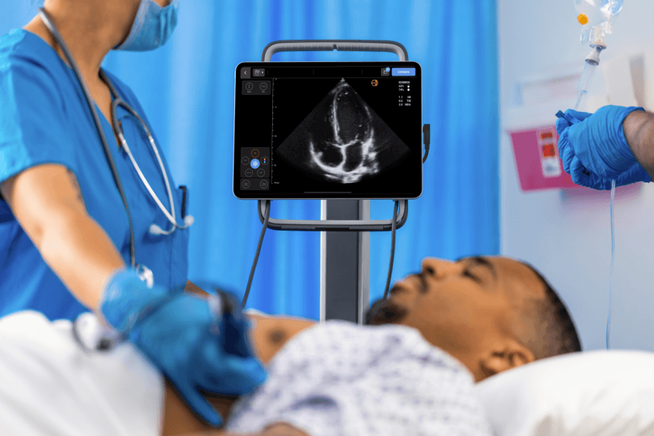
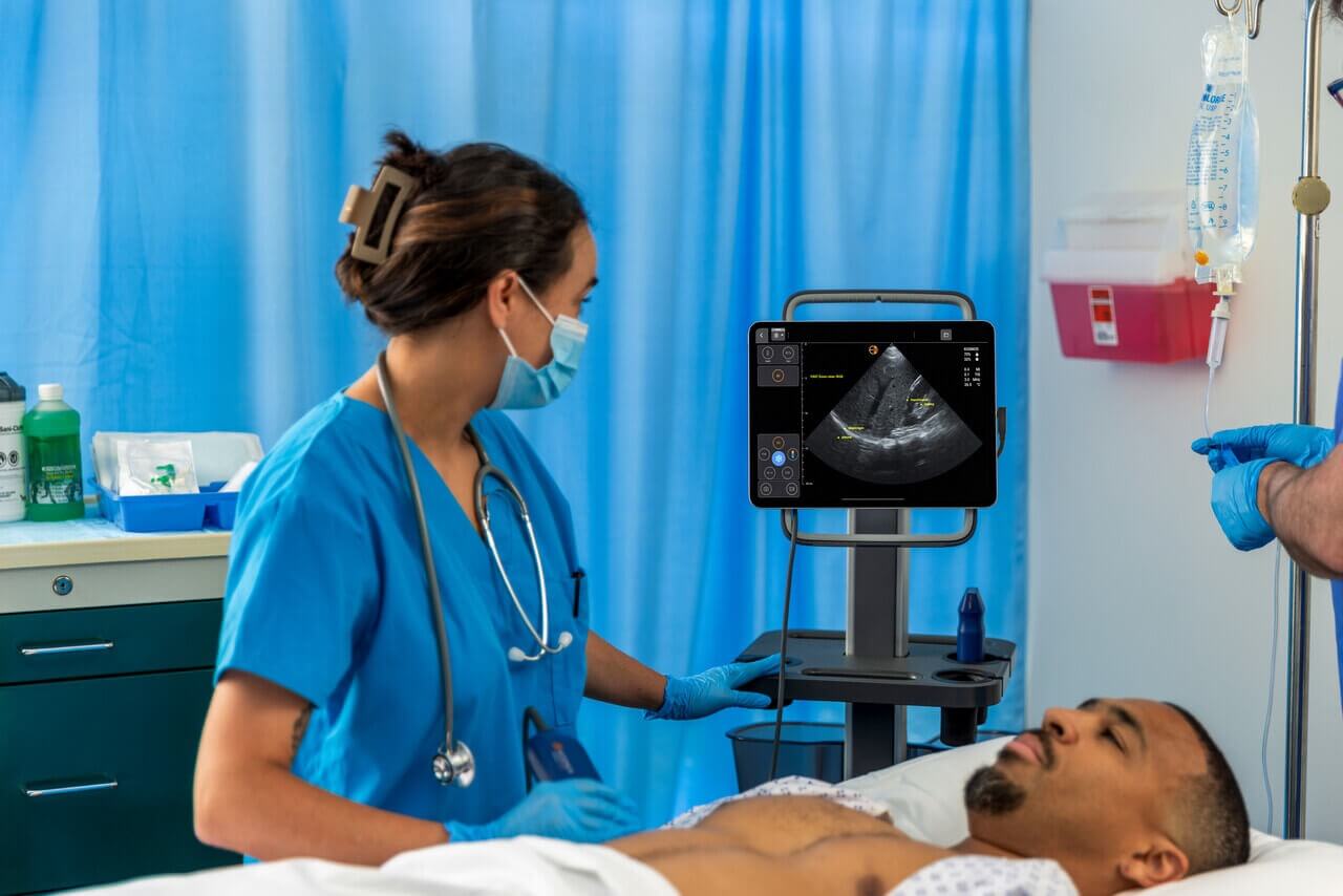
Point-of-care ultrasound (POCUS) has revolutionized bedside diagnostics, offering nurses the ability to assess bladder conditions in real-time. This non-invasive tool eliminates the need for more invasive procedures like catheterization and provides immediate results.
POCUS for Bladder Imaging
POCUS involves using portable ultrasound devices to visualize the bladder, allowing clinicians to detect abnormalities such as stones, residual urine, and structural changes. Unlike traditional imaging techniques, POCUS can be performed at the bedside with minimal preparation, making it ideal for busy clinical environments (5).
Common ultrasound Features of Bladder Stones
- Acoustic Shadowing: Stones appear as hyperechoic (bright) structures that cast dark shadows behind them. This shadowing occurs because stones block the passage of sound waves (4).
- Twinkling Artifact: On Doppler imaging, stones produce a rapidly alternating color pattern, mimicking turbulent flow. This artifact is particularly helpful in identifying small stones that may not create prominent shadows (4).
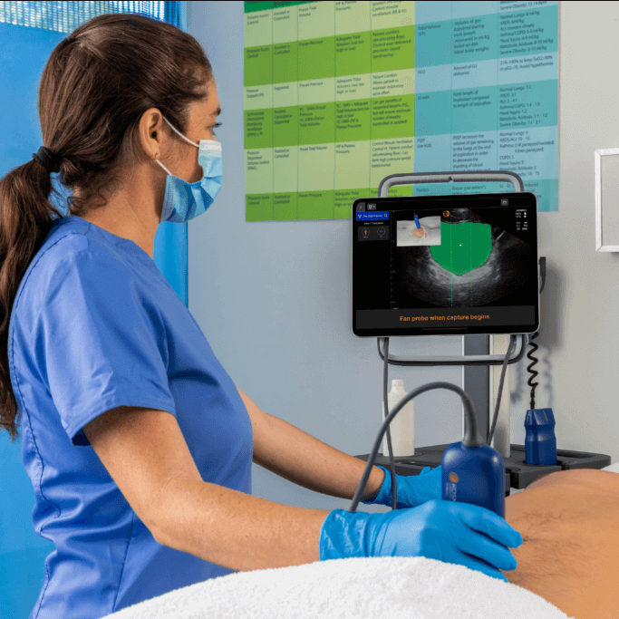
How to Perform a Bladder Ultrasound
Bladder ultrasound is an effective, non-invasive method for detecting stones and evaluating residual urine volume. Proper technique ensures accurate results and minimizes patient discomfort.
Preparing the Patient for Ultrasound
- Position the patient supine with the lower abdomen exposed.
- Explain the procedure to alleviate patient anxiety.
- Use a generous amount of ultrasound gel on the probe to improve image clarity.
Key Steps in Ultrasound Scanning
- Use a low-frequency transducer (for example, Kosmos Torso-One) to capture both transverse and longitudinal bladder views.
- Identify echogenic (bright) structures that cast posterior acoustic shadows indicative of stones.
- Adjust the patient’s position or ask them to cough to observe stone mobility.
Interpreting Ultrasound Findings
- Confirm the presence of residual urine by scanning before and after voiding.
- Detect twinkling artifacts on Doppler Imaging, which are sensitive indicators of small stones (6).
- Differentiate stones from other bladder abnormalities, such as tumors or blood clots, using multiple imaging planes.
Applications of Portable Ultrasound Equipment in Nursing
Bladder ultrasound has several practical applications in nursing practice, ranging from diagnosis of urinary retention to managing catheters (7).
Identifying Residual Urine and Retention
Bladder ultrasound helps assess post-void residual urine, a critical metric for diagnosing urinary retention. Identifying and managing retention early can prevent complications such as infections and kidney damage (1,7).
Catheter Management and Biofeedback (8,9)
- Catheter Functionality: Ultrasound can determine whether catheters are draining properly or if blockages are present.
- Biofeedback Training: Bladder retraining programs use ultrasound to provide real-time feedback to patients with urinary incontinence.
Monitoring Patients with Neurogenic Bladder
Regular bladder ultrasounds are recommended for patients with neurogenic bladder to monitor for complications such as infections, hydronephrosis, or bladder stones (5).
Conclusion and Next Steps
Bladder stones, while often preventable, can lead to severe complications if left untreated. By integrating portable ultrasound tools like POCUS into routine care, nurses can improve diagnostic accuracy and patient outcomes. Devices like Kosmos provide an accessible, non-invasive solution for detecting stones and monitoring bladder health.
To further explore the use of ultrasound in bladder imaging, access more resources or schedule a demo.
References
1. Leslie, S. W., et al. (2023). Bladder Stones. In StatPearls. StatPearls Publishing.
2. Norsworthy, A. N., et al. (2017). From Catheter to Kidney Stone: The Uropathogenic Lifestyle of Proteus mirabilis. Trends in Microbiology, 25(4), 304–315. PMID: 28017513
3. Glazer, K., et al. (2025). Ureterolithiasis. In StatPearls. StatPearls Publishing.
4. Koratala, A., et al. (2019). Bladder stone: “Must know” ultrasonographic signs. Clinical Case Reports, 7(3), 573–574. PMID: 30899498
5. Addison, R. (2000, October 5). A guide to bladder ultrasound. Nursing Times.
6. Bacha, R., et al. (2019). Clinical Significance of Twinkling Artifact in the Diagnosis of Urinary Stones. Ultrasound in Medicine & Biology, 45(12), 3199–3206. PMID: 31537388
7. Medical Advisory Secretariat. (2006). Portable bladder ultrasound: An evidence-based analysis. Ontario Health Technology Assessment Series, 6(11), 1–51. PMID: 23074481
8. Haylen, B. T., et al. (1989). Assessing the effectiveness of different urinary catheters in emptying the bladder: An application of transvaginal ultrasound. British Journal of Urology, 64(4), 353–356. PMID: 26843359. Schallom, M., et al. (2020).
9. Accuracy of Measuring Bladder Volumes with Ultrasound and Bladder Scanning in the ICU. American Journal of Critical Care : An Official Publication, American Association of Critical-Care Nurses, 29(6), 458–467. PMID: 33130866