Sonogram vs. Ultrasound: What’s the Difference?
From sound waves to screen: making sense of the terms and the tech
In the comparison of sonogram vs ultrasound, new and sometimes even existing users may seem to use the terms interchangeably. In the simplest explanation, ultrasound is the sound-wave technology and the examination, while a sonogram is an image produced during that examination. They describe different parts of the same process: the ultrasound scan creates the sonogram that a clinician uses to assess anatomy.¹
A patient says, “I’m having a sonogram.” Their appointment letter says “ultrasound.” A student watching a bedside scan hears “sonography” and “echo.” The vocabulary can make one technology sound like several different tests.
Understanding the terms helps patients follow an explanation and helps learners describe what they are seeing. The distinction also matters when discussing a complete imaging examination versus a focused bedside assessment. Here’s how the words fit together, how the images form, and where ultrasound belongs in clinical care.
Table of Contents
Sonogram vs ultrasound: the terms defined
Ultrasound refers to sound at frequencies above human hearing. In medical imaging, it also names the technology and the examination that use those waves to evaluate structures inside the body. The probe sends sound into tissue and receives returning echoes; the system processes those signals into an image.²
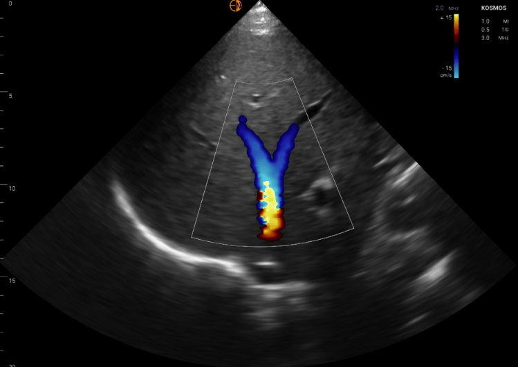
Sonogram showing hepatic veins
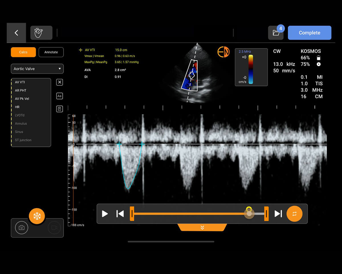
Sonogram means the resulting image. Think of the relationship as an examination and its output: the ultrasound is performed, and sonograms are captured. An examination may produce many saved images and video clips rather than one picture.¹
Sonography is another name for ultrasound imaging. A sonographer is a healthcare professional who acquires ultrasound images. Depending on the setting, another trained clinician may perform the scan.¹
An echocardiogram, often shortened to “echo,” is an ultrasound examination of the heart. Doppler ultrasound evaluates movement, commonly blood flow, and may be included within an examination.³
| Term | What it describes | Example |
|---|---|---|
| Ultrasound | Sound-wave imaging technology or the examination | An abdominal ultrasound examination |
| Sonogram | An image produced by ultrasound | A saved image of the bladder |
| Sonography | The practice of ultrasound imaging | Training in diagnostic sonography |
| Sonographer | A professional who acquires ultrasound images | A sonographer performing a scheduled scan |
| Echocardiogram | Ultrasound imaging of the heart | A study of cardiac structure and function |
| Doppler ultrasound | Assessment of movement, including blood flow | Evaluating flow within a vessel |
The practical distinction is between the method, the person performing it, and the information produced. A sonogram is part of the examination; interpretation turns imaging findings into a clinically useful assessment.
Why the terms get used interchangeably
In everyday conversation, “getting a sonogram” usually means having an ultrasound examination. Pregnancy scans make this usage especially familiar: people often focus on the image they receive rather than the equipment that created it. Both words can therefore communicate the same appointment, even though their technical meanings differ.¹
Clinicians can acknowledge a patient’s wording while explaining the test clearly: “We’ll use ultrasound to look at the area and save images for review.”
For an order or clinical record, “ultrasound” with the body region and intended examination is more useful than “sonogram” alone. A bedside report should describe the study, indication, findings, clinical impression, and important limitations. Expert consensus supports structured documentation, including documentation of technically inadequate examinations.⁴
For example, “focused bladder ultrasound to assess bladder volume” communicates more than “sonogram done.” The name should help the next reader understand what question the examination was intended to answer.
How ultrasound creates a sonogram
Image formation begins with the transducer, the probe placed against the skin. In many probes, piezoelectric materials convert electrical energy into mechanical vibrations. Those vibrations generate ultrasound waves. The same materials convert returning sound waves into electrical signals that the system can process.⁵
Gel helps sound pass between the probe and skin by reducing intervening air. As waves travel through the body, some return as echoes from tissue boundaries. The system uses the time an echo takes to return to calculate depth, then combines that information with echo strength to construct an image.⁵
The familiar grayscale picture is a map of returning echoes, rather than a photograph. Fluid often looks dark because it produces relatively few internal echoes; other interfaces appear brighter. Probe position changes the slice of anatomy shown on screen.²
Frequency creates a practical tradeoff. Higher frequencies generally improve detail in superficial structures but penetrate less deeply. Lower frequencies are useful for deeper targets. Probe selection therefore depends on what the clinician needs to examine.⁶
Three common imaging modes present different information:²,⁶
- B-mode, or brightness mode: Displays a two-dimensional grayscale view of anatomy.
- M-mode, or motion mode: Displays changes along one ultrasound line over time, helping show movement.
- Doppler: Uses changes in reflected sound frequency to assess motion, including blood flow. Color and spectral displays present that information in different ways.
Image quality also depends on settings and technique. Artifacts can obscure anatomy or create misleading appearances, so acquiring a recognizable picture is only one part of the skill. Interpretation requires understanding how the image was produced.⁶
For a closer look at the probe itself, explore EchoNous’s guide to ultrasound crystals and physics.
Where ultrasound is used
Ultrasound is used well beyond pregnancy care. Formal examinations may take place in radiology departments, cardiac imaging laboratories, outpatient clinics, or other services. A sonographer often acquires the images, and an appropriately qualified clinician interprets the examination. The workflow depends on the study and setting.¹,³
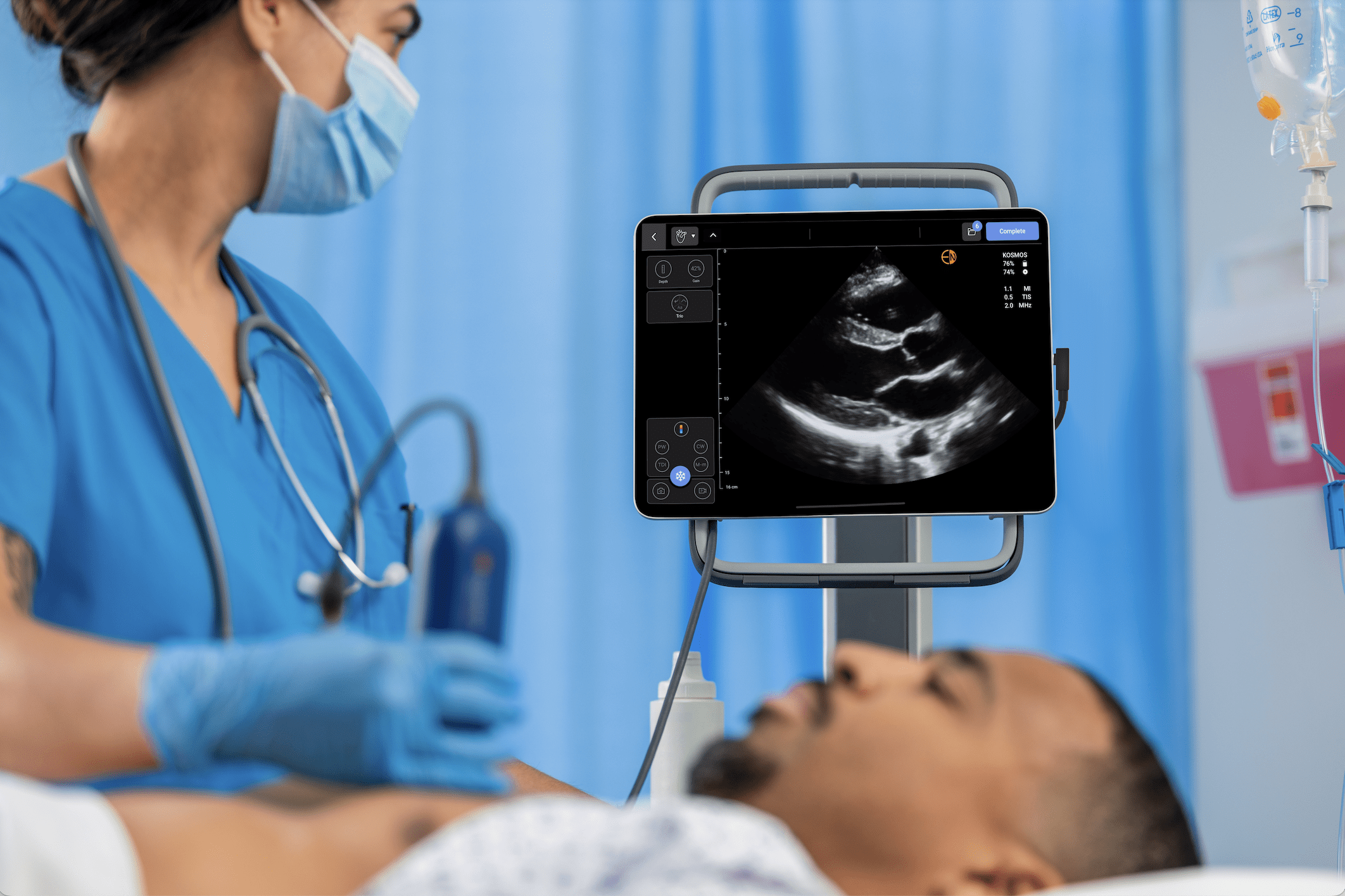
Point-of-care ultrasound (POCUS) brings image acquisition and interpretation into the treating team’s clinical assessment. It commonly addresses a focused question or supports a procedure, with findings considered alongside the history and physical examination.⁴
Examples of ultrasound applications include:³,⁵,⁷
- Cardiac: Examining heart structure, pumping function, or fluid around the heart.
- Lung: Evaluating the pleural region and fluid-related changes accessible to ultrasound.
- Abdominal: Examining organs such as the liver, gallbladder, and kidneys.
- Vascular access: Visualizing vessels to support needle placement.
- Bladder: Evaluating bladder filling or volume.
- Obstetric: Assessing pregnancy and fetal anatomy within the appropriate examination.
The setting does not change the relationship between the terms. A scheduled abdominal ultrasound and a bedside bladder scan both use ultrasound technology to produce images.
What changes is the clinical question and the scope of the examination. A focused cardiac assessment, for example, does not necessarily supply the detail of a comprehensive echocardiogram. Training, image quality, and uncertainty determine when additional imaging or expert review is needed.⁷
For learners, describing the question first is a useful habit: “What are we trying to find out?” EchoNous’s POCUS overview introduces the broader bedside workflow.
Ultrasound vs other imaging
Ultrasound provides real-time imaging without ionizing radiation. Portable equipment also allows scanning at the bedside. These properties make it useful when clinicians need to watch movement or guide a procedure.³
Other modalities answer different questions:
| Modality | How it forms images | Practical strengths and considerations |
|---|---|---|
| Ultrasound | Sound waves | Real-time views of accessible anatomy; image quality depends on the acoustic window and operator.³ |
| X-ray | Ionizing radiation | Commonly used to assess the chest and bones; overlapping structures can limit detail.⁸ |
| Computed tomography (CT) | X-rays processed into cross-sectional images | Detailed views across body regions; uses ionizing radiation and may require contrast.⁹ |
| Magnetic resonance imaging (MRI) | Magnetic fields and radiofrequency energy | Detailed soft-tissue imaging without ionizing radiation; examination time and implant compatibility matter.¹⁰ |
Ultrasound is often less expensive than CT or MRI, but actual costs depend on the examination and healthcare setting.³ Price alone does not determine the right test.
Air and bone can limit ultrasound access to deeper structures. A technically limited scan should not be treated as proof that a condition is absent.⁶ The appropriate modality depends on the clinical question, the area being examined, and whether the images provide enough information to act.
Handheld ultrasound today
Ultrasound systems now include compact and handheld options alongside larger cart-based equipment. Miniaturization allows clinicians to bring imaging into more examination rooms and bedside encounters. It also supports teaching focused scanning as part of clinical assessment.⁷
A smaller system still requires trained users. Its usefulness depends on the intended examination, available probes and imaging modes, and a workflow for reviewing and recording findings.⁷
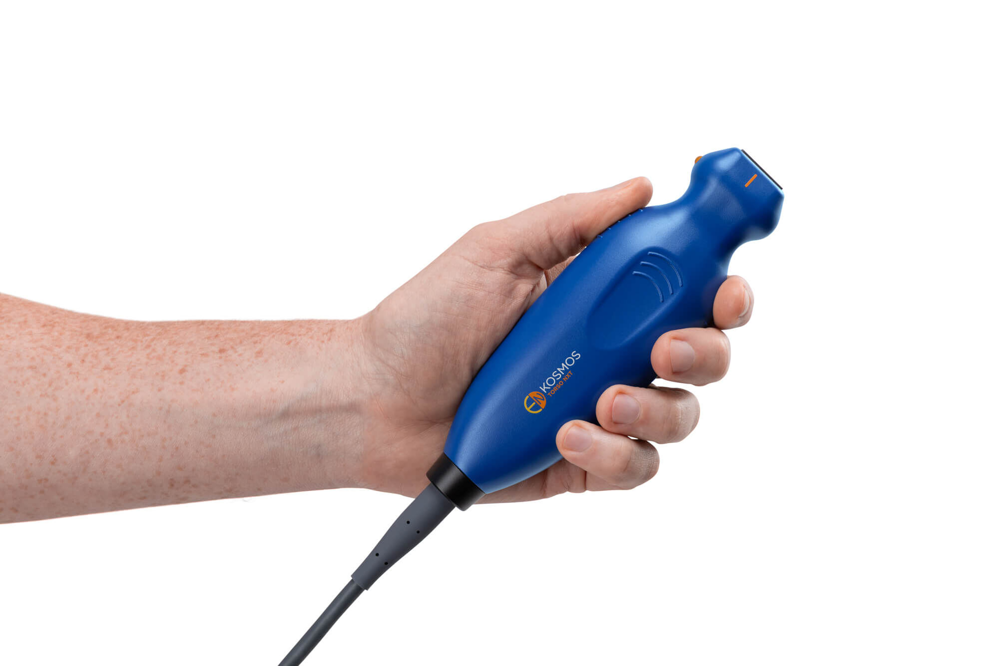
Kosmos by EchoNous is one example of a portable ultrasound platform. EchoNous’s product resources provide documentation for its configurations and software.¹¹
For learners or teams comparing equipment, EchoNous’s guide to point-of-care ultrasound device cost offers a starting point for considering the purchase. Clinical requirements, training, and ongoing use should shape that discussion alongside price.
Conclusion
Ultrasound is the technology and examination; a sonogram is an image it creates. Understanding that distinction makes conversations clearer, from a first pregnancy scan to a focused bedside assessment. Students can continue learning through EchoNous’s educational resources, including Kosmos EDU, an application designed to support ultrasound education.¹²
Questions and answers
The words often describe the same appointment in everyday conversation. Technically, ultrasound is the examination and technology; a sonogram is an image it produces.¹
No. Ultrasound also produces images of the heart, blood vessels, bladder, abdominal organs, and other structures. Pregnancy imaging is one familiar application.³
A sonographer or another appropriately trained healthcare professional performs the ultrasound examination that creates the sonogram. At the point of care, a trained clinician may acquire and interpret images within their scope of practice.¹,⁷
Diagnostic ultrasound uses sound waves, not ionizing radiation. It should still be performed for an appropriate purpose with suitable settings and examination time.⁵
An ultrasound order requests an examination of a specified body region or clinical problem. A sonogram is an image from that examination. Patients can use either word in conversation; clinical orders should identify the intended study.⁴
References
- Cleveland Clinic. Ultrasound. Accessed October 10, 2026. Source.
- Poggi C, Palavecino M. Ultrasound principles and instrumentation. Surg Open Sci. 2024;18:123-128. doi:10.1016/j.sopen.2024.02.005.
- American College of Radiology, Radiological Society of North America. General ultrasound. RadiologyInfo.org. Reviewed September 23, 2024. Accessed October 10, 2026. Source.
- Oto B, Baeten R, Chen L, et al. Best practices for point of care ultrasound: an interdisciplinary expert consensus. POCUS J. 2024;9(1):95-108. doi:10.24908/pocus.v9i1.17240.
- National Institute of Biomedical Imaging and Bioengineering. Ultrasound. Accessed October 10, 2026. Source.
- Abu-Zidan FM, Hefny AF, Corr P. Clinical ultrasound physics. J Emerg Trauma Shock. 2011;4(4):501-503. doi:10.4103/0974-2700.86646.
- Cardim N, Dalen H, Voigt JU, et al. The use of handheld ultrasound devices: a position statement of the European Association of Cardiovascular Imaging (2018 update). Eur Heart J Cardiovasc Imaging. 2019;20(3):245-252. doi:10.1093/ehjci/jey145.
- American College of Radiology, Radiological Society of North America. Chest x-ray. RadiologyInfo.org. Accessed October 10, 2026. Source.
- American College of Radiology, Radiological Society of North America. Body CT. RadiologyInfo.org. Accessed October 10, 2026. Source.
- American College of Radiology, Radiological Society of North America. Body MRI. RadiologyInfo.org. Reviewed June 15, 2026. Accessed October 10, 2026. Source.
- EchoNous. Kosmos by EchoNous resources. Accessed October 10, 2026. Source.
- EchoNous. Kosmos EDU. Accessed October 10, 2026. Source.