POCUS First for Small Bone Fractures:
A Modern, Evidence-Based Approach for Reducing X-Ray Delays and Radiation
In the emergency department (ED) or acute care setting, few things are as routine as a suspected fracture of a finger, toe, hand, or foot. These small bones—the phalanges, metacarpals, and metatarsals—are injured often, with metacarpal fractures alone accounting for 10% of all fractures1.
Yet, their small size and the critical function they serve mean that a missed or delayed diagnosis can lead to serious, long-term complications, such as osteomyelitis or complex regional pain syndrome. Traditionally, clinicians have relied on X-rays to confirm these injuries, especially since the physical exam alone is highly unreliable, with sensitivity rates as low as 25%2.
However, the time required to get a patient to the X-ray suite and then wait for image processing and interpretation can clog the ED and equate to longer wait times. Moreover, radiographs have a downside: they may miss subtle or occult fractures, especially in small bones or in pediatric patients.

This reality is prompting a major shift in how to approach these common injuries. Point-of-care ultrasound (POCUS) has emerged as a powerful, radiation-free adjunct in this context, enabling real-time fracture detection at the bedside. This benefit is particularly important for children, who are more susceptible to the cumulative, long-term risks of ionizing radiation exposure, including a small but measurable increase in lifetime cancer risk.
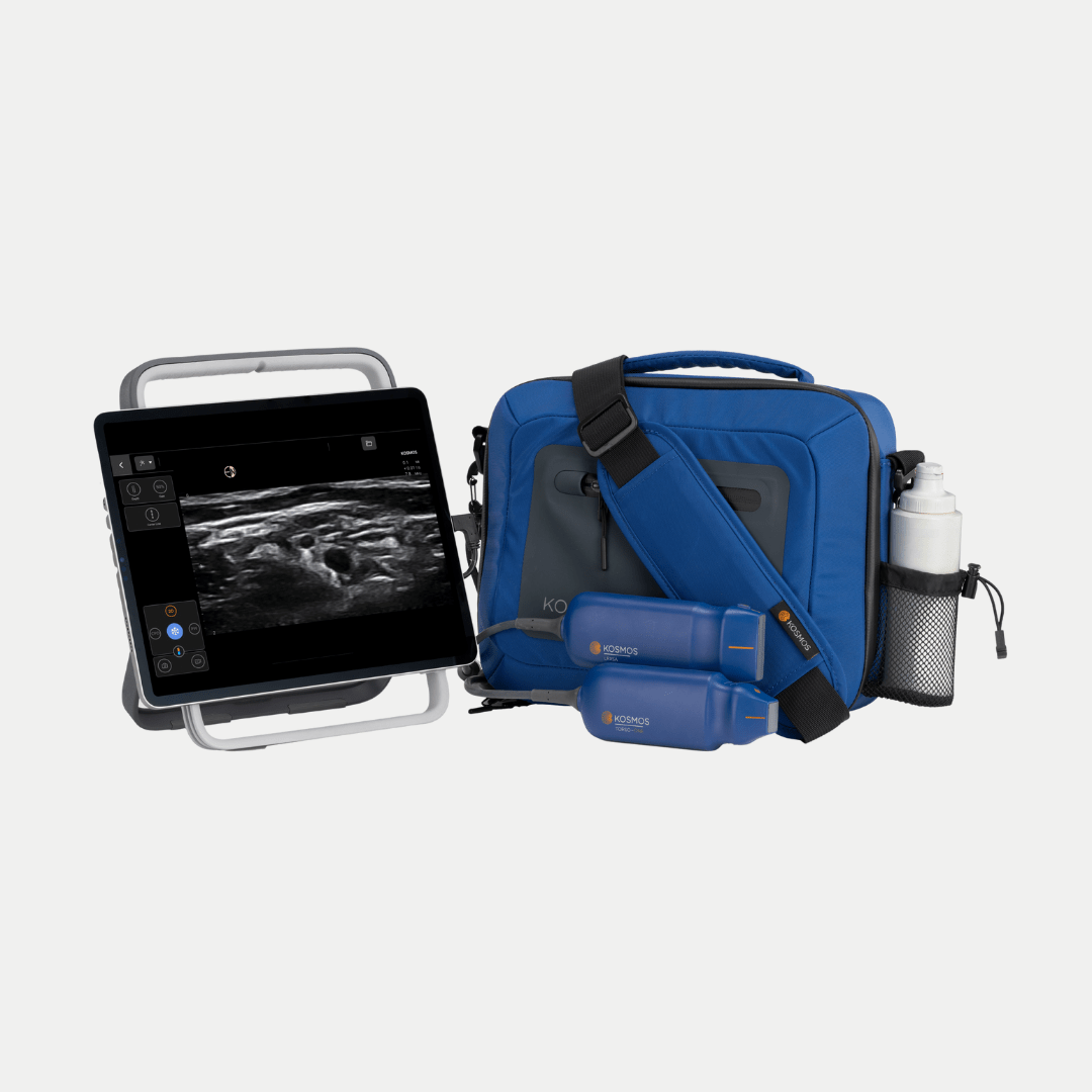
Point-of-care ultrasound solutions like the Kosmos portable ultrasound platform, with its high-frequency (up to 10.5 MHz) linear transducer, the Kosmos Lexsa, are now leading the charge in high-resolution musculoskeletal imaging.
Table of Contents
- POCUS First for Small Bone Fractures:
- A Modern, Evidence-Based Approach for Reducing X-Ray Delays and Radiation
- Limitations of Radiographs in Small Bone Fractures
- Ultrasound and Technique for Fracture Detection “High-Value Hybrid” Tier
- Ultrasound Technique with the Lexsa Probe
- Integrating POCUS Into Fracture Evaluation
- Q&A: POCUS for Small Bone Fractures
- References
Limitations of Radiographs in Small Bone Fractures
Conventional radiography remains the first-line imaging modality for suspected fractures, but it has important limitations in the acute ED setting. Small, non-displaced fractures in the fingers or toes can be radiographically occult or difficult to discern on initial X-rays. Overlapping anatomy and the need for perfect positioning can lead to missed injuries.
For instance, certain foot injuries (e.g. subtle metatarsal fractures or Lisfranc injuries) are notoriously easy to overlook on plain films. In pediatric patients, growth plates and cartilaginous components may obscure or mimic fractures on X-ray, complicating interpretation.
Further, in the post SARS-CoV-2 pandemic era, with concerns of ED boarding and ED flow times, obtaining X-rays means that a patient must be transported to radiography or have a radiology technologist perform a portable study, then wait for image processing and interpretation, equating to longer wait times.3 Ultimately, while the radiograph may be the gold standard, especially to help demonstrate angulation and similar characteristics, POCUS is well versed to rule out any hand or foot fractures in the ED, with sensitivity and specificity of close to 100%. 4, 5
Ultrasound and Technique for Fracture Detection “High-Value Hybrid” Tier
By allowing the clinician to visualize the bone at the bedside, POCUS can enable immediate correlation with the site of pain or deformity within seconds 6 and subsequent treatment or discharge.
Importantly, ultrasound can detect fractures by revealing cortical disruptions that indicate a break. When a high-frequency linear probe like the Lexsa is placed over a bone, the normal cortex appears as a continuous hyperechoic (bright) line with acoustic shadowing beyond.
A fracture manifests as an interruption or step-off in that bright line. Because ultrasound beams cannot penetrate intact bone cortex, any discontinuity allows some sound to pass into the bone, often creating an acoustic artifact (an “echoic wedge”) at the break.
Clinically, this appears as a gap or irregularity in the smooth cortical line. Surrounding features such as a localized hematoma or periosteal elevation may also be visualized as hypoechoic (dark) collections adjacent to the fracture site, further supporting the diagnosis.
Using the Lexsa probe’s high-resolution capabilities, even hairline fractures can be identified if the transducer is carefully aligned along the bone. In children, ultrasound has the added benefit of visualizing cartilaginous structures; it can delineate buckle (torus) or greenstick fractures of immature bone that might be subtle on X-ray. 7 Ultimately, the evidence is clear that POCUS can identify even occult fractures missed on radiography with minimal false positives. 5, 8
Ultrasound Technique with the Lexsa Probe
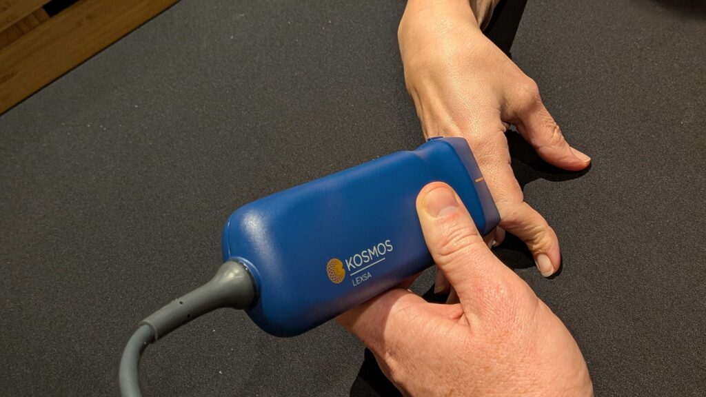
Proper technique is key to maximizing ultrasound’s diagnostic yield for fractures. The EchoNous Lexsa linear array probe (64/128 channels) is designed for high-frequency, high-resolution imaging of superficial structures.
The probe is placed in a longitudinal orientation along the length of the bone. Scanning the area of maximal tenderness, the clinician sweeps the probe through the region, carefully inspecting the bony cortex on the ultrasound screen.
A normal cortex appears as a smooth echogenic line; the examiner should look for any disruption in this line. Even a small step-off of the cortical surface is suggestive of a fracture. It is important to scan in two orthogonal planes, i.e. longitudinal (long-axis) and transverse (short-axis) to fully appreciate the extent and orientation of a fracture, and to avoid missing small cracks that might only be visible in one plane. Gentle pressure with the probe can elicit pain directly over a hidden fracture (the “tenderness localization” sign), aiding in targeting the correct area.
For very superficial injuries (such as distal fingertip fractures), a thick gel standoff or water bath technique can be used to improve near-field focus, though the Lexsa probe’s minimum scanning depth of 1 cm should be sufficient for fingers and toes.
Using the probe’s high-frequency settings (3–10.5 MHz range, 7.5 MHz Center Frequency) provides optimal axial resolution to visualize tiny cortical defects. The clinician should compare the injured side with the contralateral side if uncertainty arises (in pediatric cases, this can help differentiate growth plate from fracture line). Through practice, one becomes adept at recognizing the hallmark ultrasound findings of fracture, i.e. cortical break, shadow interruption, and peri-fracture hematoma.
Integrating POCUS Into Fracture Evaluation
The advent of high-fidelity POCUS devices like the Kosmos Lexsa by EchoNous calls for a reassessment of the traditional fracture workup in the ED. The goal is not to eliminate X-rays, but to use ultrasound to triage and expedite care. A modern, evidence-based approach might be as follows:
- Clinical Assessment: Evaluate the injured extremity for deformity, swelling, focal bony tenderness, and functional impairment. Apply decision rules (e.g. Ottawa Ankle/Foot Rules for foot injuries) to gauge pre-test probability of fracture.
- POCUS First: If a fracture is suspected in a readily accessible location (phalanges, metacarpals, metatarsals), perform a bedside ultrasound with a high-frequency linear probe (Lexsa) as an initial diagnostic step. Focus on the area of maximal tenderness.
- Interpretation:
- Positive Ultrasound (Fracture Identified): Confirm the presence of a cortical break or step-off on ultrasound, which is diagnostic of fracture. Proceed with appropriate management (immobilization, splinting, pain control). In many cases, a confirmatory X-ray can be obtained subsequently for documentation and to assess fracture details (e.g. degree of displacement), however, the treatment/consults can be initiated immediately, after the ultrasound result. For non-operatively managed fractures (e.g. a simple toe fracture), POCUS findings may potentially obviate the need for any radiograph, especially if follow-up outpatient X-rays can be arranged. 9
- Negative Ultrasound (No Fracture Seen): If no cortical disruption is seen and the patient’s exam is otherwise reassuring (mild pain, able to bear weight or move the finger, etc.), the likelihood of a significant fracture is very low. The injury can be managed conservatively (e.g. buddy taping a sprained finger) with instructions for outpatient follow-up. Here, POCUS’s high negative predictive value helps avoid unnecessary radiographs and ED length of stay.5
- Equivocal Ultrasound or High Clinical Suspicion: If the ultrasound exam is limited (due to pain, swelling, or anatomic difficulty) or if clinical suspicion for fracture remains high despite a negative scan, then proceed with standard radiography or other imaging. POCUS should augment but not completely replace clinical judgment; certain complex injuries (e.g. intra-articular fractures, open fractures or fractures with subtle alignment issues) may still require X-ray/CT for full evaluation.
By prioritizing POCUS for suitable cases, clinicians can reduce time to diagnosis, minimize radiation exposure, and allocate X-ray resources to where they are most needed. Not every fracture evaluation will start with ultrasound, but as the technology (and familiarity with it) grows, more will and evidence suggests many patients stand to benefit.
Q&A: POCUS for Small Bone Fractures
POCUS offers real-time, bedside imaging that can detect cortical disruptions with near-100% sensitivity and specificity. It reduces delays associated with X-rays and eliminates radiation exposure—making it ideal for fingers, toes, hand, and foot injuries.
Yes. Evidence shows POCUS can detect even subtle, hairline, or occult fractures—many of which may be missed on radiographs, especially in small bones or pediatric patients.
Small, superficial fractures such as those in the phalanges, metacarpals, and metatarsals. POCUS is especially helpful for non-displaced, occult, and pediatric fractures.
A high-frequency linear probe is recommended. The Kosmos Lexsa (3–10.5 MHz, 7.5MHz Center Frequency) provides exceptional resolution for visualizing cortical irregularities and peri-fracture findings.
The key ultrasound signs of a fracture involve disruptions to the bone’s cortex, primarily manifesting as a break or step-off in the bright, continuous cortical line; this discontinuity is often accompanied by an “echoic wedge” or artifact at the site of the injury, and may also be suggested by the presence of a localized hematoma or periosteal elevation.
Not entirely. While POCUS can diagnose many fractures and speed up clinical decision-making, X-rays may still be needed for documentation, assessing displacement, or evaluating complex or intra-articular injuries.
By scanning at the bedside, clinicians can rapidly diagnose or rule out fractures, reducing transport delays, decreasing ED length of stay, and prioritizing X-ray resources for higher-acuity cases.
References
- Bernstein ML, Chung KC. Hand fractures and their management: an international view. Injury. Nov 2006; 37(11):1043-1048. doi:10.1016/j.injury.2006.07.020
- Krastman P, Mathijssen NM, Bierma-Zeinstra SMA, et al. Diagnostic accuracy of history taking, physical examination and imaging for phalangeal, metacarpal and carpal fractures: a systematic review update. BMC Musculoskeletal Disorders. 2020; 21(1)doi:10.1186/s12891-019-2988-z
- Osterwalder J, Hoffmann B, Blaivas M, et al. A Plea for a Paradigm Shift from X-Ray to Ultrasound in Adults: An Update for Emergency Physicians, General Practitioners, Orthopedists and Sports Medicine Physicians. Diagnostics (Basel). Jul 21 2025; 15(14)doi:10.3390/diagnostics15141827
- Perlmutter JW, Olajide E, Elsherbini A, et al. Integrating Point-of-Care Ultrasound in Hand Clinic: A Systematic Review and Meta-Analysis. J Hand Surg Glob Online. Sep 2025; 7(5):100775. doi:10.1016/j.jhsg.2025.100775
- Champagne N, Eadie L, Regan L, et al. The effectiveness of ultrasound in the detection of fractures in adults with suspected upper or lower limb injury: a systematic review and subgroup meta-analysis. BMC Emerg Med. Jan 28 2019; 19(1):17. doi:10.1186/s12873-019-0226-5
- Poonai N, Myslik F, Joubert G, et al. Point-of-care Ultrasound for Nonangulated Distal Forearm Fractures in Children: Test Performance Characteristics and Patient-centered Outcomes. Acad Emerg Med. May 2017; 24(5):607-616. doi:10.1111/acem.13146
- Cocco G, Ricci V, Villani M, et al. Ultrasound imaging of bone fractures. Insights into Imaging. 2022; 13(1)doi:10.1186/s13244-022-01335-z
- Hoffman DF, Adams E, Bianchi S. Ultrasonography of fractures in sports medicine. British Journal of Sports Medicine. 2015; 49(3):152-160. doi:10.1136/bjsports-2014-0942179.
- Gorbachova T, Chang EY, Ha AS, et al. ACR Appropriateness Criteria® Acute Trauma to the Foot. Journal of the American College of Radiology. 2020; 17(5):S2-S11. doi:10.1016/j.jacr.2020.01.019