Revolutionizing Aortic Stenosis Diagnosis: Dr. Jon Zubiaur Zamacola on Handheld Ultrasound Innovation

Dr. Jon Zubiaur Zamacola
Cardiovascular diseases remain one of the leading causes of death worldwide, and aortic stenosis (AS) is a particularly significant condition affecting millions of patients. Early diagnosis and continuous monitoring are essential to prevent severe complications, yet access to high-end echocardiography systems is often limited in many clinical settings.
Dr. Jon Zubiaur Zamacola, an Interventional Cardiology Fellow at Hospital Universitario La Paz in Madrid, Spain, has dedicated his research to improving bedside cardiac imaging. His recent study explores the potential of handheld ultrasound devices, particularly the Kosmos ultrasound system, in assessing aortic stenosis at the point of care.
“As a cardiologist, I wanted to explore whether a handheld ultrasound device could provide the necessary accuracy to assess aortic stenosis severity without needing a full echocardiography lab,” explains Dr. Zubiaur.
This raises an important question: can handheld ultrasound technology provide a reliable alternative to traditional echocardiography for diagnosing and monitoring aortic stenosis? In this article, we delve into Dr. Zubiaur’s research, its clinical implications, and the future of portable cardiac ultrasound in modern healthcare.
The Growing Need for Portable Cardiac Ultrasound Challenges in Aortic Stenosis Diagnosis
Challenges in Aortic Stenosis Diagnosis
Aortic stenosis (AS) is a progressive heart valve disease that restricts blood flow from the heart, leading to serious complications such as heart failure, arrhythmias, and sudden cardiac death [1]. Timely and accurate diagnosis is critical, yet the current standard for AS evaluation relies on expensive high-end echocardiography systems, requires skilled operators, and is not always readily available, particularly in rural or resource-limited healthcare settings [2,3].
Dr. Jon Zubiaur Zamacola highlights this issue: “In many hospitals, access to high-end echocardiography is limited, and transporting critically ill patients to imaging labs can be challenging. A portable solution could bridge this gap and allow immediate bedside assessment.”
The Global Impact of Heart Valve Disease
Heart valve diseases, including aortic stenosis, affect over 100 million people worldwide [4,5]. With aging populations, the prevalence of AS is rising rapidly, particularly in developed countries, where degenerative calcification of the aortic valve is standard [5]. In low-resource settings, patients often go undiagnosed or untreated due to a lack of access to specialized cardiac imaging [4].
The Role of Portable Ultrasound in Expanding Access
Handheld ultrasound technology offers a promising solution to these accessibility challenges. Unlike traditional echocardiography machines, portable ultrasound devices like Kosmos by EchoNous provide [6]:
- Bedside diagnostics, reducing the need to transport critically ill patients
- Lower-cost alternatives to high-end echocardiography
- Potential use in primary care and remote settings for early AS detection
Dr. Zubiaur emphasizes the potential of handheld ultrasound in modern cardiology:
“We are entering an era where portable ultrasound could become an essential tool, especially in settings where access to comprehensive echocardiography is limited. This could be a game-changer for early and more frequent monitoring of aortic stenosis patients.”
By improving access to diagnostic tools, handheld ultrasound can revolutionize how aortic stenosis is diagnosed and managed, particularly in underserved regions and point-of-care settings [7–9].
Why Dr. Zubiaur Conducted This Study
A Passion for Improving Bedside Diagnostics
Dr. Jon Zubiaur Zamacola has always been drawn to innovative approaches in cardiology, particularly those that can improve patient care at the bedside. As an Interventional Cardiology fellow at Hospital Universitario La Paz in Madrid, his work revolves around advanced cardiac imaging, valve disease, and catheter-based interventions.
His passion for bedside diagnostics developed during his early training, where he observed the challenges clinicians faced in quickly assessing cardiac conditions without immediate access to a full echocardiography suite. This inspired him to explore portable ultrasound as a potential solution for real-time cardiac evaluation.
“In cardiology, imaging is everything. The ability to assess aortic stenosis severity at the bedside, without needing a large ultrasound system, could change the way we practice, “ says Dr. Zubiaur.
Identifying Gaps in Traditional Ultrasound
Despite significant advances in cardiac imaging, traditional echocardiography systems remain:
- Expensive, often costing hundreds of thousands of dollars
- Bulky and immobile, limiting their use in emergency rooms, ICUs, and remote clinics
- Operator-dependent, requiring highly skilled personnel to achieve assessments
Many portable ultrasound devices lack key features for comprehensive aortic stenosis evaluation, such as continuous wave Doppler (CW) – a critical tool for measuring the severity of blood flow obstruction through the aortic valve [9]. Recognizing this gap, Dr. Zubiaur sought to validate a handheld device that could rival high-end echocardiography systems in diagnosing aortic stenosis.
The Importance of Continuous Wave Doppler
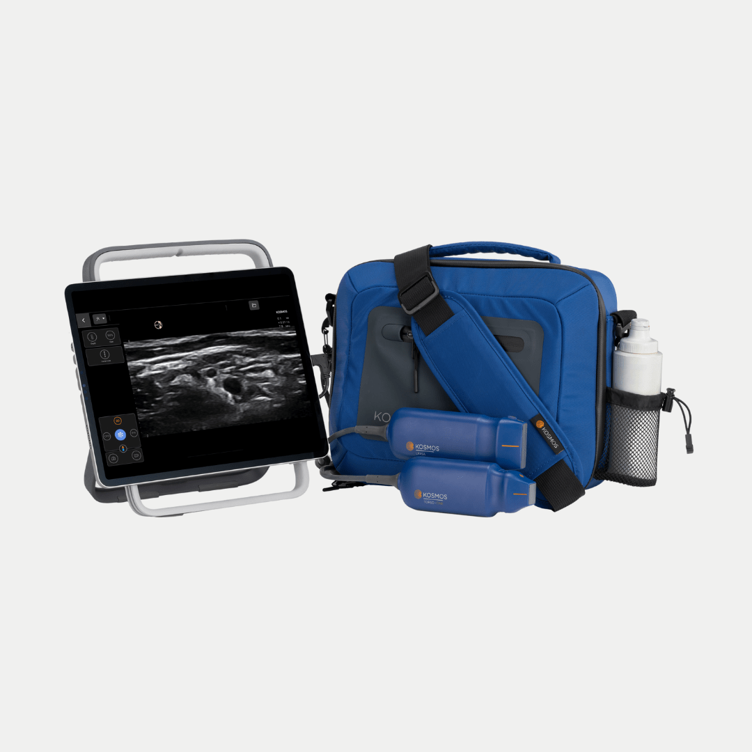
Dr. Zubiaur and his team selected the Kosmos ultrasound device by EchoNous for their study because it was one of the only handheld ultrasound systems with continuous wave Doppler capability. This feature is essential for:
- Measuring the aortic valve velocity is the primary metric for assessing stenosis severity
- It provides real-time, accurate blood flow analysis similar to high-end machines
- Enabling rapid bedside assessment without needing full laboratory echocardiography
“Kosmos stood out because it was the first portable device we found that allowed us to perform continuous wave Doppler measurements that were previously only available in full-sized echocardiography machines,” explains Dr. Zubiaur.
Study Design and Key Findings[9]
Methodology
Dr. Jon Zubiaur Zamacola and his team conducted a prospective validation study to evaluate the accuracy of the Kosmos handheld ultrasound device compared to a high-end echocardiography system for assessing aortic stenosis.
The study involved 101 patients with suspected aortic stenosis, who underwent:
- Besides assessment using Kosmos handheld ultrasound
- Standard echocardiography with a high-end machine
To ensure accuracy and eliminate bias, the study used a blinded methodology, where the clinicians interpreting the Kosmos results were unaware of the findings from the high-end system and vice versa.
“We designed this study to reflect real-world clinical settings. Our goal was to see whether a handheld device could provide the same level of diagnostic reliability as a traditional echocardiography machine, “explains Dr. Zubiaur.
The primary measurement was the mean aortic valve gradient, a critical factor in determining stenosis severity.
Key Results
Strong Agreement with High-End Echocardiography
- Kosmos performed comparably to a full-scale echocardiography machine in measuring mean aortic valve gradients, confirming strong diagnostic accuracy.
- Over 90% accuracy in detecting severe aortic stenosis, making it a reliable bedside screening tool.
“We were very encouraged by the level of agreement between Kosmos and the standard echocardiography system. This validates its potential as an accessible, point-of-care diagnostic tool,” says Dr. Zubiaur.
Challenges and Areas for Improvement
- Obese patients showed reduced image quality, affecting Doppler signal acquisition
- Atrial Fibrillation (Afib) patients had variability in gradient measurements, requiring additional refinements in signal processing
These findings confirm that Kosmos is a powerful tool for rapid cardiac evaluation, but further technological enhancements could improve its performance in complex cases.
Clinical Implications for Cardiology
Transforming Emergency & Primary Care Workflows
The ability to rapidly assess aortic stenosis at the bedside could significantly improve:
- Triage in emergency rooms, ensuring early diagnosis and intervention
- Primary care screenings, allowing earlier detection and referral for at-risk patients
- ICU and inpatient monitoring, reducing the need for costly and time-consuming transfers to imaging labs
“With handheld ultrasound, we can assess a patient in real-time rather than waiting for an echocardiography appointment. This speeds up decision-making and improves patient outcomes,” Dr. Zubiaur explains.
Cost Comparison: Traditional vs. Handheld Ultrasound
- High-end echocardiography systems: $100,000+, require dedicated lab space and highly trained specialists.
- Kosmos handheld ultrasound: Significantly more affordable, portable, and easy to use, reducing healthcare costs and increasing accessibility.
Future Applications
- Broader adoption in outpatient cardiology clinics
- Use in global health initiatives to expand access to cardiac diagnostics
- Integration with telemedicine, allowing specialists to guide remote evaluations
Handheld ultrasound technology is poised to revolutionize cardiac diagnostics, ensuring earlier detection and better patient management.
Addressing Limitations & Future Innovations
Why Obese & AFib Patients Present Challenges
While Kosmos performed well overall, two patient groups showed notable limitations:
- Obese patients: Reduced image clarity due to increased tissue depth made Doppler measurements more challenging
- Atrial fibrillation patients: The irregular heartbeat affected gradient measurements, requiring refinements in Doppler waveform processing
“These limitations highlight the need for further refinement in portable ultrasound technology, particularly for more complex patient populations,” notes Dr. Zubiaur.
AI & Software Advancements to Improve Performance
Advances in AI-driven ultrasound technology could enhance accuracy and reduce variability in difficult cases.
- AI-guided Doppler alignment for better readings in obese patients
- Automated rhythm correction to improve gradient calculations in AFib patients
- Machine learning algorithms that optimize ultrasound settings in real-time
“As AI integration continues, handheld ultrasound will only become more powerful, making it an indispensable tool in cardiac diagnostics,” predicts Dr. Zubiaur.
With ongoing technological advancements, handheld ultrasound is set to become a mainstream tool, bridging the gap between accessibility, affordability, and diagnostic precision in modern cardiology.
Conclusion
The ability to diagnose and monitor aortic stenosis quickly and accurately is critical in modern cardiology. Dr. Jon Zubiaur Zamacola’s research has shown that handheld ultrasound devices like Kosmos offer a reliable, accessible, and cost-effective alternative to high-end echocardiography.
With over 90% accuracy in detecting severe aortic stenosis, Kosmos can be integrated into emergency rooms, primary care clinics, and resource-limited settings, ensuring earlier diagnosis and better patient outcomes.
“Handheld ultrasound is no longer just a convenience—it’s a necessity. With the right technology, we can improve access to cardiac care for millions of patients worldwide,” says Dr. Zubiaur.
As the healthcare industry moves toward more portable and AI-driven diagnostics, handheld ultrasound is set to play a key role in the future of cardiology.
Want to learn more? Explore the benefits of Kosmos for your practice. Request a demo today and take your cardiac diagnostics to the next level.
Are you ready to revolutionize your practice with portable ultrasound technology?
Schedule a demo today and explore how Kosmos can transform cardiac assessments in your clinical setting.
Citations
1. Pujari, S. H., et al. (2025). Aortic Stenosis. In StatPearls. StatPearls Publishing.
2. Fazlalizadeh, H., et al. (2024). Closing the “Last Mile Gap” in Access to Multimodality Imaging in Rural Settings: Design of The Imaging Core of The Risk Underlying Rural Areas Longitudinal Study. Circulation. Cardiovascular Imaging, 17(2), e015496.
3. Chamsi-Pasha, M. A., et al. (2017). Handheld Echocardiography. Circulation, 136(22), 2178–2188.
4. Chen, Q., et al. (2024). Global, Regional, and National Burden of Valvular Heart Disease, 1990 to 2021. Journal of the American Heart Association, 13(24), e037991.
5. Lindman, B. R., et al. (2016). Calcific aortic stenosis. Nature Reviews. Disease Primers, 2, 16006.
6. Perez-Sanchez, A., et al. (2024). Comparison of 6 handheld ultrasound devices by point-of-care ultrasound experts: A cross-sectional study. The Ultrasound Journal, 16(1), 45.
7. Venkatayogi, N., et al. (2023). From Seeing to Knowing with Artificial Intelligence: A Scoping Review of Point-of-Care Ultrasound in Low-Resource Settings. Applied Sciences, 13(14), Article 14.
8. Wen, S., et al. (2023). Point-of-Care Ultrasound in Detection, Severity and Mechanism of Significant Valvular Heart Disease and Clinical Management. Journal of Clinical Medicine, 12(20), 6474.
9. Zubiaur, J., et al. (2025). Validation of a hand-held ultrasound device in the evaluation of aortic stenosis. The International Journal of Cardiovascular Imaging, 41(2), 377–385.