Ultrasound for Monitoring Diaphragm Dysfunction after Cardiac Surgery
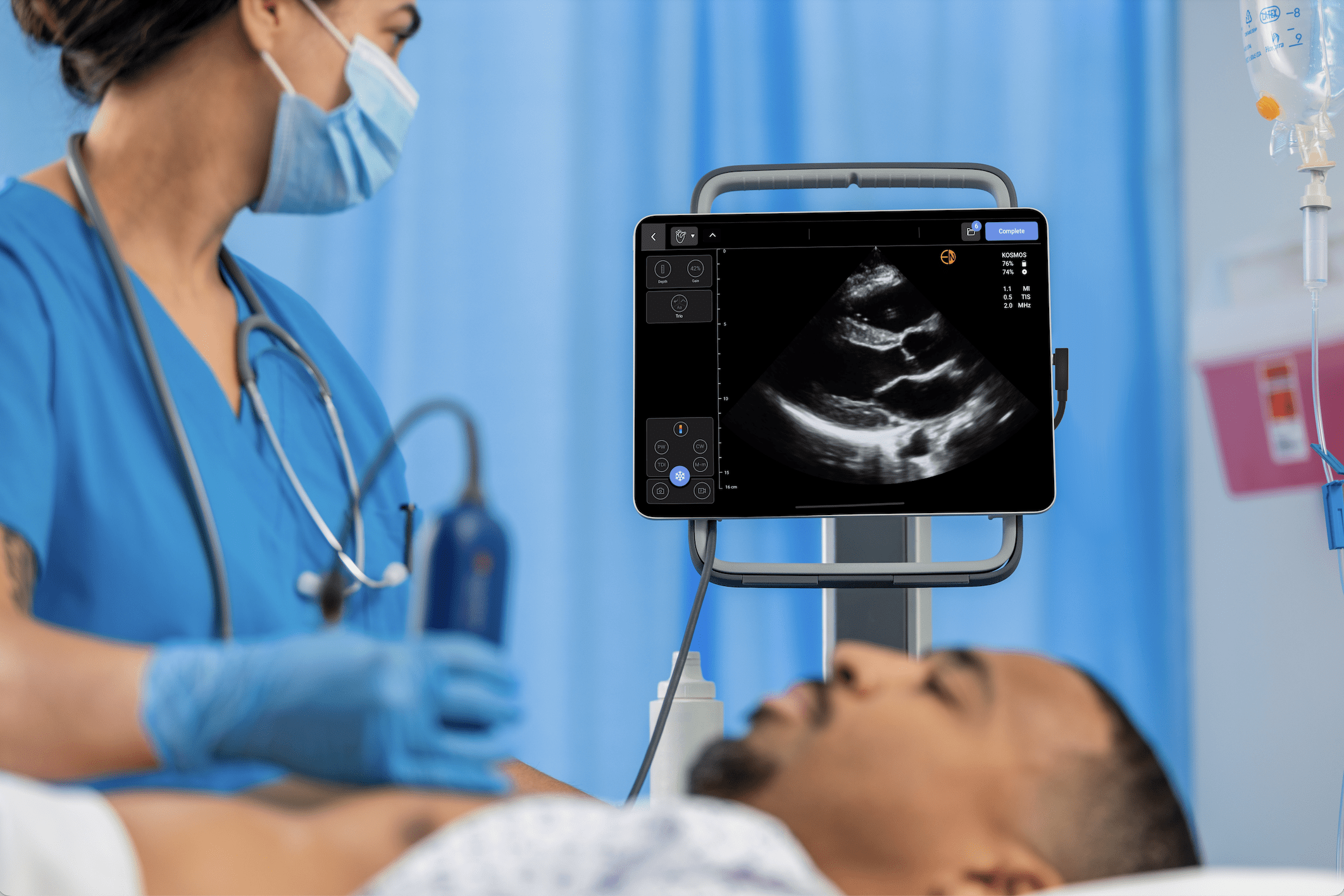
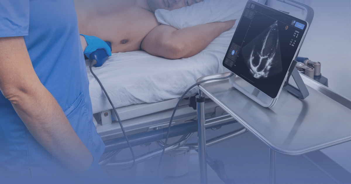
Point-of-care ultrasound (POCUS) is gaining a reputation as a bedside diagnostic and monitoring tool in critical patient care. This reputation is mostly connected to its effectiveness, versatility, and less invasive approach to patient assessment.
Recently, POCUS has been deployed to estimate respiratory function in critically ill patients. Ultrasound modalities exist for estimating post-surgery respiratory function, assessing the post-operative integrity of abdominal muscles, and tracking the clinical progression of diaphragm dysfunction.
Considering its wide applications in cardiology today, this article reviews available clinical evidence supporting point-of-care ultrasound modalities in investigating the diaphragm function during the recovery phase of cardiac surgery.
Read about ultrasound applications in critical care here.
Key Takeaways
- The utility of point-of-care ultrasound (POCUS) is expanding to include post-surgery patient monitoring in cardiology and pulmonology
- With its innervations originating from the central nervous systems and crisscrossing around the cardiovascular system, the diaphragm is perhaps the most important physiological connection among the cardiovascular, central nervous, and respiratory systems
- Observational studies have reported diaphragm dysfunction cases – paresis, weakness, and paralysis – during the recovery phase of patients who had undergone cardiac surgery; in most cases, the diaphragm nerves or its fibro-muscular structure is damaged
- Ultrasound modalities provide a valid and reliable assessment of diagram integrity, helping clinicians accurately track and manage cases of diaphragmatic dysfunction in critically ill, post-operative patients
Table of contents
Ultrasound for Assessing Diaphragmatic Dysfunction
In 2011, an observational study published in the Journal of Critical Care Medicine kicked off the adoption of ultrasound for assessing diaphragmatic dysfunction. A group of scientists from Ulsal University College of Medicine, Seoul, Korea, trialed M-mode ultrasonography to measure vertical excursion or paradoxical movements indicating diaphragmatic dysfunction in critically ill patients.
Study results showed how the ultrasound technique accurately diagnosed a substantial number of diaphragmatic dysfunctions linked with early and delayed treatment failures. Like others, this study backed ultrasound assessment of diaphragmatic integrity in critically ill patients who had undergone cardiac surgery (1).
Now, clinicians readily perform routine evaluations after cardiac surgery to ascertain the diaphragm’s functional status. These evaluations investigate two diaphragm sonographic properties: the diaphragmatic excursion (DE), a measurement of the distance the diaphragm moves during a respiratory cycle, and the diaphragm thickening factor (DTF), a variation in the diaphragm’s thickness during a respiratory effort (2).
Diaphragm Integrity in Post-Cardiac Surgery Recovery & Clinical Consequences
The diaphragm, the major respiratory muscle, is a dome-shaped, fibro-muscular structure composed of a central tendon shielded by peripheral muscle fibers. With an excursion of 1 – 2 cm and a forced breathing amplitude of up to 11 cm, the diaphragm is responsible for about 60 – 80% of the respiratory workload.
In its anatomical makeup, the diaphragm has two hemidiaphragms with the right one positioned slightly higher than the left. As a fibro-muscular structure, the diaphragm is innervated by two phrenic nerves originating from the C3 – C5 nerve roots – a connection bridge between the respiratory and the central nervous systems. Both phrenic nerve groups navigate the neck, thorax, and the mediastinal surface of the parietal plural to connect with each hemidiaphragm.
Physiological Integrity
In addition to the central nervous system, the nerve connections of the hemidiaphragm also maintain close connections with another important organ system: the cardiovascular system.
The left phrenic nerve is close to the subclavian artery and passes in front of the pericardial sac of the left ventricle; the right phrenic nerve runs superficially to the anterior scalene muscle and the subclavian artery and passes over the right atrium and right ventricle.
Vessels from the intercostal arteries and collaterals from the abdominal aorta and the internal mammary artery collectively supply arterial blood flow to the diaphragm (3).
Functionally, the diaphragm’s close connection to the cardiovascular system (as described) posed a major problem in cardiac surgeries – procedures and tools deployed in heart surgeries – like nerve repair, artery bypass, and angioplasty – pose serious structural and functional damages to the hemidiaphragm. In fact, cases like these are commonly observed after atrial fibrillation ablation (1).
Clinical Consequence
Phrenic nerve injuries resulting from direct surgical trauma or indirect injury (due to stretching by the sternal retractor) significantly impair diaphragmatic function. In serious cases, a complete loss of one hemidiaphragm induces a restrictive syndrome with a resultant 25% decrease in vital capacity.
A recent observational study re-investigating the incidence, risk factor, and clinical consequence of diaphragmatic dysfunction adds more context – the researchers reported a 7.6% incidence rate of diaphragmatic dysfunction leading to serious respiratory complications and prolonged ICU stays (4).
Ultrasound Modalities for Estimating Diaphragm Integrity
Ultrasound analysis of diaphragm function is becoming more important in cardiology – after all, the frequency of diaphragm dysfunction is significant after cardiac surgery. Today, clinical studies focus on two primary use cases. These include:
a. Estimating Diaphragm Mobility
Diagrammatic ultrasound modality helps cardiologists and other clinicians visualize and evaluate the diaphragm’s mobility through three principal views: the anterior subcostal, posterior subcostal, and subxiphoid views. Using a two-dimensional (B) mode or one-dimensional (M) mode, these views measure diaphragmatic mobility at three time points; during quiet breathing, maximal inspiration, and voluntary sniffing.
In a sitting position, the posterior subcostal view is recommended – a low-frequency convex transducer over the posterior subcostal region in B mode projects a fine image of both hemidiaphragm through the hepatic or spleen windows. To measure diaphragm mobility, the amplitude of craniocaudal diaphragmatic mobility is measured in M mode. In slender adults and children, the subxiphoid view is recommended.
As in the posterior subcostal view, the transducer is positioned below the xiphoid process in a transverse orientation and angled cranially and dorsally toward the posterior hemidiaphragm. Motility is measured by evaluating the excursion of each hemidiaphragm.
b. Diagnosing Hemidiaphragm Paralysis
Tracking hemidiaphragm activity is important in post-cardiac surgery patients. Diaphragmatic paralysis can arise from either a weakness of the muscles or severe damage to the diaphragm innervations.
In patients with a clinical indication of diaphragmatic paralysis, hemidiaphragm movement is either paradoxical or completely absent at rest. Diaphragm ultrasound – using M-mode ultrasonography – detects a paradoxical cranial movement of the hemidiaphragm in these patients.
At maximal inspiration, the first paradoxical movement is characterized by a biphasic movement and a cranio-caudal excursion. These observations are combined with data on inspiratory thickening to decide on hemidiaphragm paralysis.
In 2023, the International Journal of Health Sciences published a study investigating ultrasound accuracy in the diaphragm motion assessment as a clinical predictor of diaphragm paralysis (5).
The study gathered laboratory investigations and imaging results from 163 pediatric patients and compared the diagnostic accuracies of ultrasound, chest X-ray, laboratory investigations, and clinical suspicion. Ultrasound modalities yielded 96.6% and 97.7% sensitivity and specificity rates respectively. Compared with X-ray, clinical suspicion, and laboratory results, the data points demonstrated how ultrasonography modalities present a valid and reliable diagnosis of hemidiaphragm paralysis.
What Next?
Point-of-care ultrasound is a fast-evolving diagnostic tool. Beyond its diagnostic imaging use cases in cardiac care, it also aids the clinical assessment of diaphragmatic function – especially in post-operative and intensive care units where patients’ cooperation is not easily obtained. With its non-radiating, less-invasive, and low-cost properties, diaphragm ultrasound should be considered a standard of care modality in the global drive to manage diaphragm dysfunction in post-operative patients.
Article by Dr. Adil Maqbool, MBBS, FCPS
Reviewed by Dr. Danielle Kelvas, MD
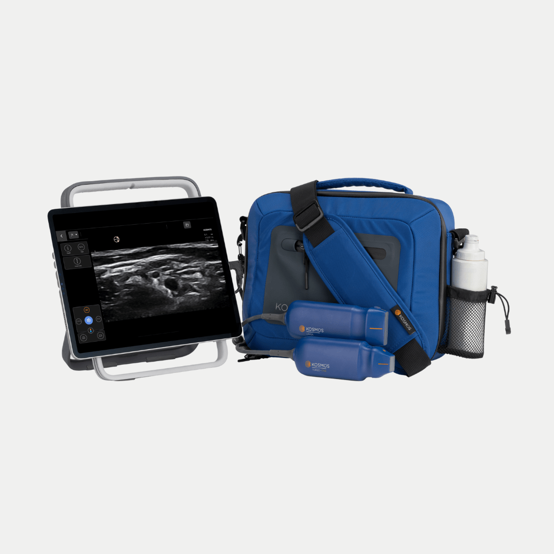
Check out Kosmos AI and Kosmos Ultraportable Ultrasound and find out more about how critical care physicians are using POCUS today
References
- Kim, W. Y., Suh, H. J., Hong, S. B., Koh, Y., & Lim, C. M. (2011). Diaphragm dysfunction assessed by ultrasonography: influence on weaning from mechanical ventilation. Critical care medicine, 39(12), 2627–2630. https://doi.org/10.1097/CCM.0b013e3182266408
- Marques, M. R., Pereira, J. M., Paiva, J. A., de Casasola-Sánchez, G. G., & Tung-Chen, Y. (2024). Ultrasonography to Access Diaphragm Dysfunction and Predict the Success of Mechanical Ventilation Weaning in Critical Care: A Narrative Review. Journal of ultrasound in medicine: official journal of the American Institute of Ultrasound in Medicine, 43(2), 223–236. https://doi.org/10.1002/jum.16363
- Boussuges, M., Blanc, P., Bregeon, F., & Boussuges, A. (2024). Interest in thoracic ultrasound after cardiac surgery or interventional cardiology. World journal of cardiology, 16(3), 118–125. https://doi.org/10.4330/wjc.v16.i3.118
- Laghlam, D., Lê, M. P., Srour, A., Monsonego, R., Estagnasié, P., Brusset, A., & Squara, P. (2021). Diaphragm Dysfunction After Cardiac Surgery: Reappraisal. Journal of cardiothoracic and vascular anesthesia, 35(11), 3241–3247. https://doi.org/10.1053/j.jvca.2021.02.023
- Aljibali A. S. (2023). Ultrasound utilization in the diagnosis of diaphragm dysfunction compared to other modalities: A retrospective study. International journal of health sciences, 17(3), 11–17.