What’s Under the Hood of Your Ultrasound Device?
Point-of-care ultrasound (POCUS) has revolutionized bedside diagnostics, allowing clinicians to visualize structures and perform interventions with precision. Portable ultrasound equipment combines advanced technologies, including harmonic imaging and compound imaging, to provide exceptional image clarity and procedural accuracy.
Table of contents
The Science Behind Kosmos POCUS Technology
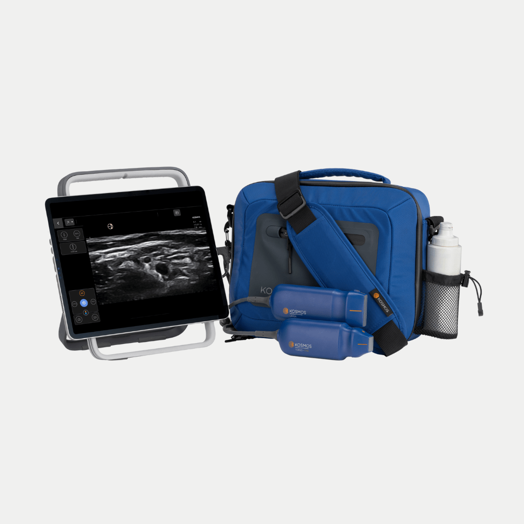
Kosmos portable ultrasound equipment integrates state-of-the-art features to enhance diagnostic accuracy and clinical workflows. It is designed to provide healthcare providers with reliable imaging solutions for diverse applications, including vascular access, cardiac assessments, bladder volume, abdominal assessment, musculoskeletal imaging, and more.
Harmonic Imaging: Improving Clarity in Diagnostics
Harmonic imaging is a fundamental advancement in ultrasound technology. Unlike conventional imaging that relies on fundamental sound wave frequencies, harmonic imaging captures second-order sound waves generated by tissue interactions. These harmonics are less susceptible to interference and noise, resulting in cleaner, sharper images [1].
Benefits of Harmonic Imaging with Kosmos POCUS:
- Noise Reduction: Reduces artifacts such as side lobes and reverberations, which can obscure critical structures.
- Enhanced Resolution: Allows for clear differentiation of soft tissues and vascular pathways.
- Improved Contrast: Distinguishes between tissues with similar acoustic properties, such as veins and arteries.
For vascular access, harmonic imaging ensures better visualization of small veins and helps clinicians perform safer, more accurate procedures.
Compound Imaging: Redefining Precision
Compound imaging takes diagnostic accuracy a step further by combining multiple frames captured from different angles into one cohesive image. This process minimizes distortions caused by refraction, shadowing, or noise, enhancing the overall clarity and reliability of the image [2].
Advantages of Compound Imaging in Ultrasound [3]:
- Minimized Shadowing: Reduces artifacts caused by dense structures such as bones or calcifications.
- Sharper Boundaries: Enhances visualization of vessel walls, making it easier to assess vascular health.
- Better Context: Provides a more comprehensive view of complex anatomical structures.
By integrating multiple frames from different angles, compound imaging reduces artifacts and enhances the accuracy of ultrasound-based diagnostics. This approach is particularly useful for visualizing complex anatomical regions, such as nerve clusters or vascular bifurcations.
Key Features of Ultrasound Equipment
Imaging Modes for Comprehensive Care
There are a variety of imaging modes that support diverse clinical applications, including:
- B-Mode: The standard grayscale imaging mode used for assessing soft tissues and anatomical structures.
- Color Doppler: Enables real-time visualization of blood flow, distinguishing arteries from veins.
- Pulsed-Wave (PW) Doppler: Measures the velocity of blood flow, aiding in cardiac and vascular assessments.
- M-Mode: Captures motion in a single scan line, making it ideal for evaluating dynamic processes such as cardiac activity.
- Continuous Wave (CW) Doppler: Allows interrogation of high-velocity jets and valvular pathology. Kosmos is the only device in its class with CW Doppler.
These imaging modes make ultrasound suitable for vascular, musculoskeletal (MSK), cardiac, and lung imaging.
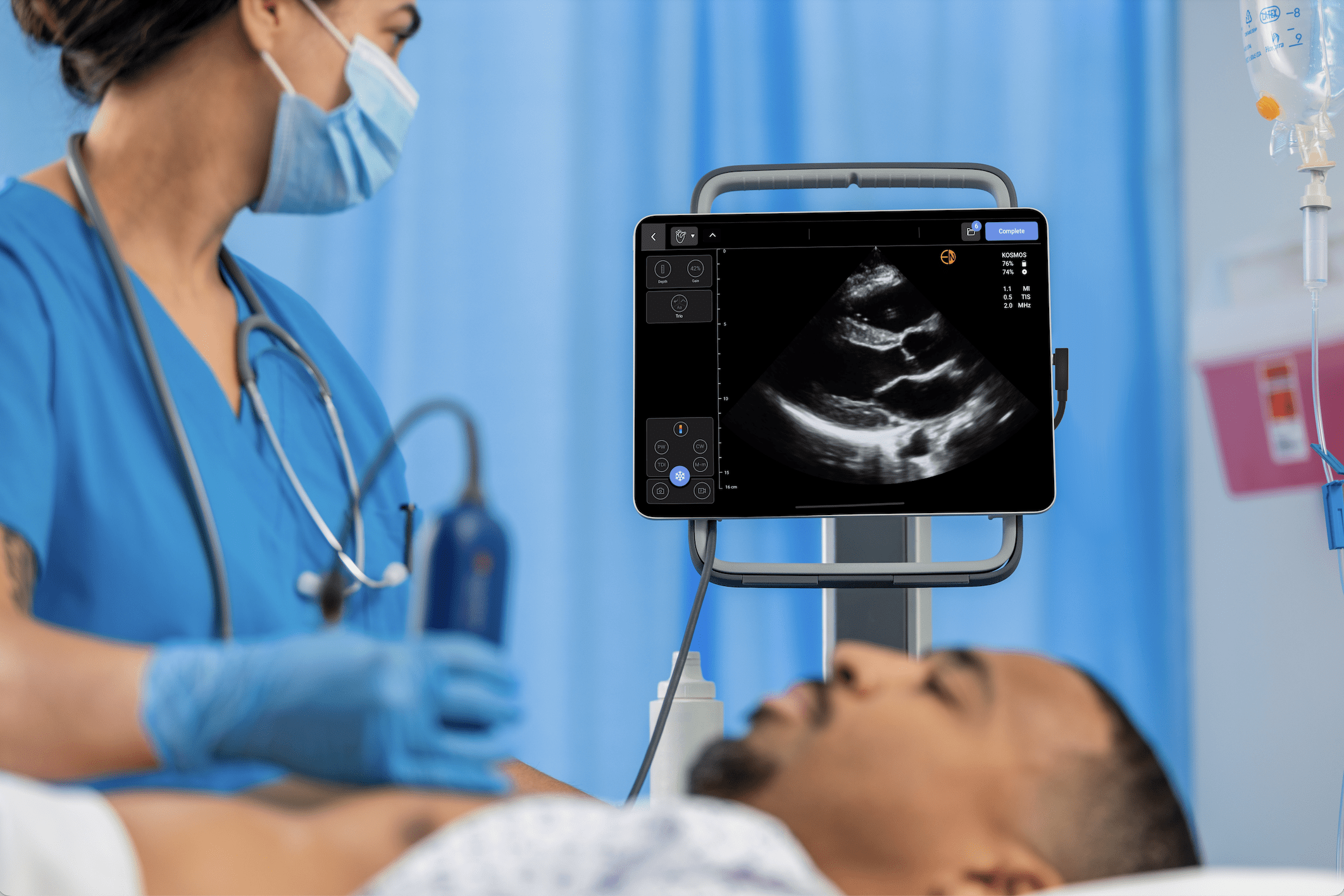
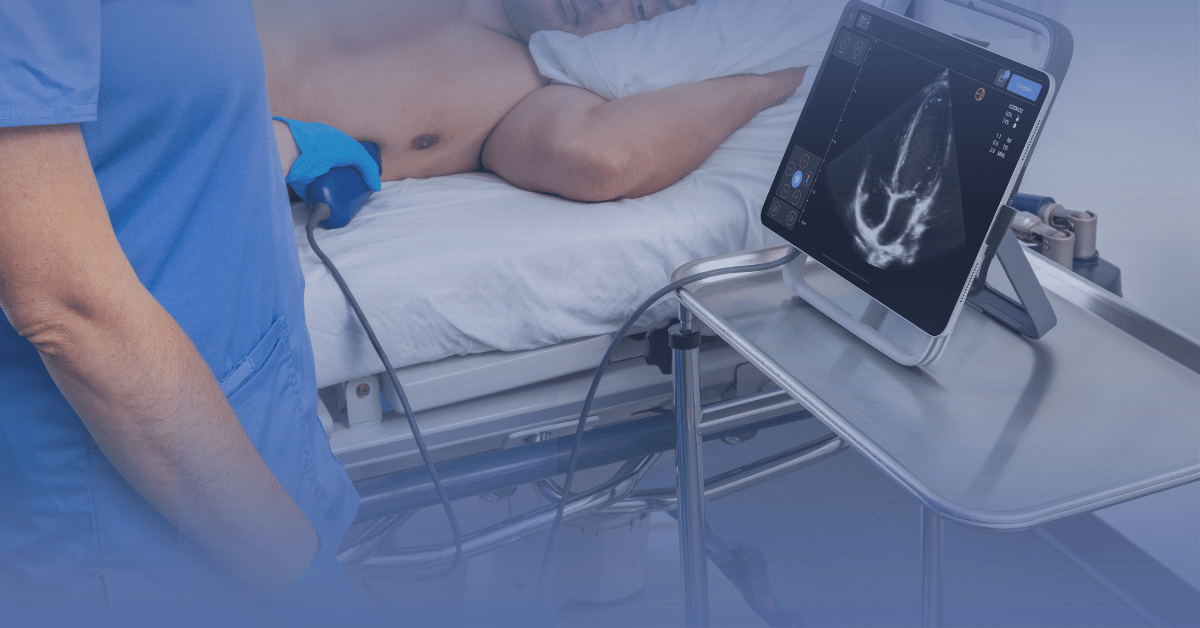
Portable Design for Point-of-Care Ultrasound
Portability is a defining feature of POCUS. Its lightweight design and user-friendly interface make it ideal for bedside use.
Key Kosmos Design Features:
- Lightweight and Compact: Torso-One and Lexsa are 275 and 280 grams respectively, making for easy handling and transport.
- Touchscreen Controls: Provides intuitive access to imaging adjustments and tools.
- Battery Life: Up to 8 hours of scanning with Kosmos Link, ensuring reliability during long procedures.
These features make Kosmos portable ultrasound equipment ideal for dynamic clinical settings, from emergency rooms to outpatient clinics.
Durability and Safety for Clinical Use
Durability is essential for devices used in fast-paced and demanding environments. Kosmos is built to withstand the rigors of clinical practice.
Durability Highlights:
- IPX7-Rated: The device is water resistant and can endure routine cleaning and disinfection.
- Drop-Tested: Designed to remain functional even after drops of up to 1 meter.
- Rapid Boot-Up: Starts in under 20 seconds, ensuring minimal downtime in critical situations.
These durability features ensure Kosmos maintains peak performance in any clinical environment.
Discover how Kosmos portable ultrasound equipment can transform your diagnostic and interventional practices. Explore its innovative features today.
Conclusion
Kosmos portable ultrasound equipment exemplifies the integration of cutting-edge POCUS technology, including harmonic imaging, compound imaging, and needle visualization. These features empower clinicians to achieve precision diagnostics and improve patient safety across various clinical settings.
References
1. Uppal, T. (2010). Tissue harmonic imaging. Australasian Journal of Ultrasound in Medicine, 13(2), 29–31.
2. Hung, A. L. Y., et al. (2021). Good and bad boundaries in ultrasound compounding: Preserving anatomic boundaries while suppressing artifacts. International Journal of Computer Assisted Radiology and Surgery, 16(11), 1957–1968. PMID: 34357525
3. Heng, H. G., et al. (2010). Appearance of common ultrasound artifacts in conventional vs. Spatial compound imaging. Veterinary Radiology & Ultrasound: The Official Journal of the American College of Veterinary Radiology and the International Veterinary Radiology Association, 51(6), 621–627. PMID: 21158234
4. Reusz, G., et al. (2014). Needle-related ultrasound artifacts and their importance in anesthetic practice. British Journal of Anaesthesia, 112(5), 794–802.
5. Peabody, C. R., et al. (2017). Deep Needle Procedures: Improving Safety With Ultrasound Visualization. Journal of Patient Safety, 13(2), 103–108.