CASA Protocol for Ultrasound Use During CPR
Advanced Life Support Protocols in Modern Healthcare
Advanced life support protocols are rapidly evolving; and with it, the different branches of modern medicine. Cardiology, nephrology, and intensive care lead the pack. The integrations of advanced life support protocols in Cardiology are particularly interesting. In recent years, researchers have published hundreds of studies evaluating the direct impacts of these integrations on patient outcomes and disease prognosis. These studies largely point to a common conclusion: advanced life support systems are critical interventions in modern medicine.
Key Takeaways
- The CASA protocol uses ultrasound during CPR to identify treatable causes of cardiac arrest and improve survival rates.
- It has three steps: check for pericardial effusion, assess right ventricular strain, and evaluate cardiac activity.
- Ultrasound helps minimize CPR interruptions by streamlining diagnosis and guiding interventions.
- Using adjunct techniques like pre-pause echo setup and quick reviews during CPR enhances effectiveness.
- Proper application of CASA with POCUS improves emergency care and patient outcomes during cardiac arrests.
Table of contents
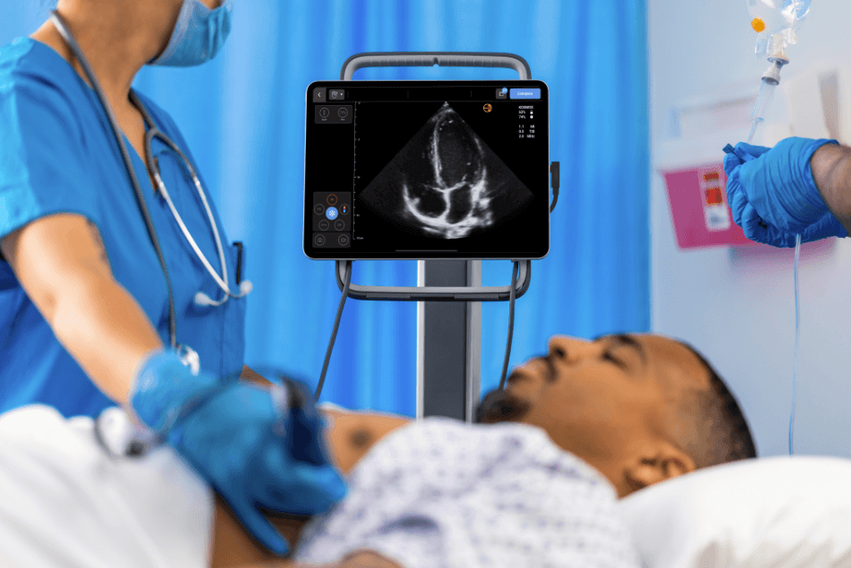
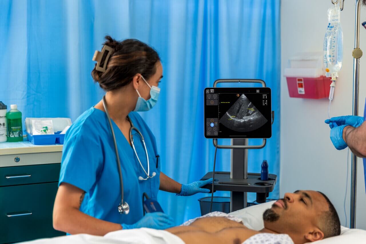
POCUS in Cardiology
Point of care ultrasound (POCUS) is used frequently in emergency and intensive care departments. Outpatient, ambulatory, and in-patient clinical settings leverage POCUS for both diagnostics and vital monitoring.
Next-gen and AI-enabled imaging technologies, as deployed in POCUS, offer pixel-perfect visualization of the cardiovascular architecture – not just in viewing how the heart pumps, but also to optimize the intricacies of vascular access in cardiac surgery (1). In modern medical practice, its clinical adoption covers the evaluation of major cardiac pathologies including pulmonary embolism, congestive heart failure, valve disease, and cardiac arrest.
Now, beyond its conventionally adopted usage, POCUS is fast gaining acceptance in a different aspect of cardiology: cardiopulmonary resuscitation. Cardiac arrest contributes large numbers to the annual global mortality figures.
In the United States alone, an estimated 356,000 out-of-hospital cardiac arrest cases are reported annually. About 90 percent are fatal, with the survival at hospital discharge pegged at 10.4%, and survival with good functional status pegged at 8.4% (2).
Annually, in-hospital cardiac arrest estimates stand at 209,000 in the United States, with an average survival ratio of 24.8%, and survival to discharge from hospital ratio of 11.4% (2). The stats for both clinical settings affirm a simple conclusion: there is an urgent need to optimize emergency cardiac care practices including cardiac resuscitation. An effective cardiac resuscitation combines high-quality cardiopulmonary resuscitation with early defibrillation for defibrillate rhythms in an attempt to end cardiopulmonary arrest.
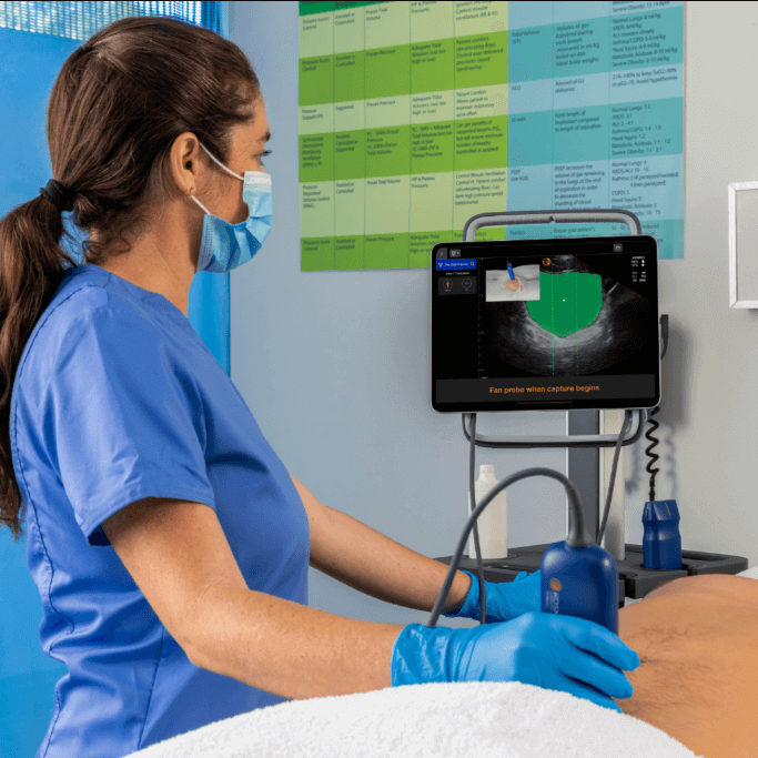
Optimizing Cardiopulmonary Resuscitation with POCUS
Portable ultrasound protocols change the game of cardiac resuscitation in modern cardiology. As an advanced life support protocol, POCUS accomplishes three primary objectives in cardiopulmonary arrest:
- Provides information on the etiology of cardiac arrest in non-defibrillate rhythms
- Assess the quality of cardiopulmonary resuscitation compressions
- Offer a data-driven prognosis of survival
In a 2018 publication, researchers from Highland Hospital-Alameda Health System Oakland assessed the effectiveness of the cardiac sonographic assessment (CASA) in reducing the duration of interruption in CPD during resuscitation. Using data collected from 267 sequential cardiac arrests, this pre versus post-intervention study demonstrated how point-of-care ultrasound significantly reduced the duration of CPR interruptions (3).
The 3-step Cardiac Arrest Sonographic Assessment (CASA) Protocol
CASA simplifies the complexities associated with CPR monitoring and assessment. Designed to govern the integration of POCUS in the resuscitation of critically ill patients, CASA facilitates the detection of reversible causes of out-of-hospital cardiac arrest (OHCA) and prevents prolonged CPR interruptions (4).
The CASA protocol, in its 3-step design, evaluates for
- Pericardial effusion
- Right heart strain
- Cardiac activity
A. Look for a pericardial effusion on the first pause
CASA starts with determining the presence of pericardial effusion – a common cause of cardiac tamponade. This evaluation informs the clinician of the immediate cause of the arrest, opening a window for possible emergency intervention. In cardiac tamponade, a pericardiocentesis may resolve pericardial effusion and positively impact prognosis early.
Patients with pericardial effusion secondary to cardiac tamponade have a better treatment outlook compared to patients with other primary causes. Although simple in theory, diagnosing a cardiac tamponade can be difficult even for the most skillful clinician. In many cases, clinicians easily miss the subtle clinical signs during initial resuscitation (5).
If tamponade is diagnosed, the decision to perform an urgent intervention must be based on ultra-sonographic findings and the patient’s clinical presentation.
B. Look for a Right ventricular (RV) Strain on the Second Pause
Right heart strain is implicated in about 4 – 8% of cardiac arrest cases. Consequently, the second step of CASA accounts for checking right heart strains as a possible trigger of pericardial effusion. Compared to other etiologies, cardiac arrests secondary to right heart strains have significant survival rates.
In this step, the clinician checks for signs of an enlarged right ventricle: a probable indication of pericardial effusion. However, the presence of an enlarged ventricle is not exclusive to pericardial effusion; chronic right ventricle disease, dysfunctional cardiac flow, and other cardiac pathologies may present with an enlarged ventricle.
If right heart strain is diagnosed, management goals should shift to attaining a return of spontaneous circulation (ROSC), followed by a detailed imaging examination with POCUS or Computed tomography.
C. Evaluate Cardiac Activity
Evaluating cardiac activity provides prognostic insight into survival chances. In patients with pericardial effusion, lack of cardiac activity moves survival to hospital discharge probability to the 0 – 0 0.6% range. Depending on practice conventions, the clinical interpretation of cardiac activity may vary. To streamline this step and properly evaluate cardiac activity, the CASA protocols recommend several rounds of cardiac compressions. Combined with available clinical and medical information, several compressions help the clinician determine the heart’s activity rate.
CASA Adjuncts
In special population patients, an optimized CASA protocol may significantly boost the survival rate. The American College of Emergency Physicians and other practice watchdogs recommend simple adjuncts in these cases. If needed, CASA adjuncts ensure high-quality PCR when using POCUS.
CASA adjuncts commonly recommended for clinicians include:
- Find the echo view before the pause
- Use a 10-second clock
- Review clip during CPR
- Stay off the chest if there are no signs of pericardial effusion or right ventricle strain
Conclusion
Beyond optimizing imaging examinations in cardiology, POCUS offers limitless potential to improve patients’ outcomes. In cardiac arrests, POCUS modalities directly assess the reversible causes of sustained interruptions in cardiac activity. If properly administered, POCUS-enabled CASA reduces clinical errors and minimizes CPR interruptions. Based on available research submissions, POCUS implementation in CASA optimizes the framework for high-quality PCR and ultimately improves the survival rate.
References
- Maheshwari, S., & Dagor, H. (2024). Evolving the Scope of Cardiac Point-of-Care Ultrasound in the Current Era. Cureus, 16(2), e53985. https://doi.org/10.7759/cureus.53985
- AHA Releases Latest Statistics on Sudden Cardiac Arrest. Available online at: https://www.sca-aware.org/sca-news/aha-releases-latest-statistics-on-sudden-cardiac-arrest. Accessed Nov 27, 2024
- Clattenburg, E. J., Wroe, P. C., Gardner, K., Schultz, C., Gelber, J., Singh, A., & Nagdev, A. (2018). Implementation of the Cardiac Arrest Sonographic Assessment (CASA) protocol for patients with cardiac arrest is associated with shorter CPR pulse checks. Resuscitation, 131, 69–73. https://doi.org/10.1016/j.resuscitation.2018.07.030
- Al Hazmi, A. (2021). Cardiac Arrest Sonographic Assessment (CASA). In: Salerno, A., Haase, D.J., Murthi, S.B. (eds) Atlas of Critical Care Echocardiography. Springer, Cham. https://doi.org/10.1007/978-3-030-74687-2_21
- Willner, D. A., Goyal, A., Grigorova, Y., Sharma, S., & Kiel, J. (2024). Pericardial Effusion. In StatPearls. StatPearls Publishing. https://pubmed.ncbi.nlm.nih.gov/28613741/